Review
. 2008 Nov;45(5):617-24.
doi: 10.1016/j.yjmcc.2008.08.001. Epub 2008 Aug 8.

The Purkinje cell; 2008 style

Affiliations
Review

The Purkinje cell; 2008 style

Wen Dun et al. J Mol Cell Cardiol. 2008 Nov.

Abstract

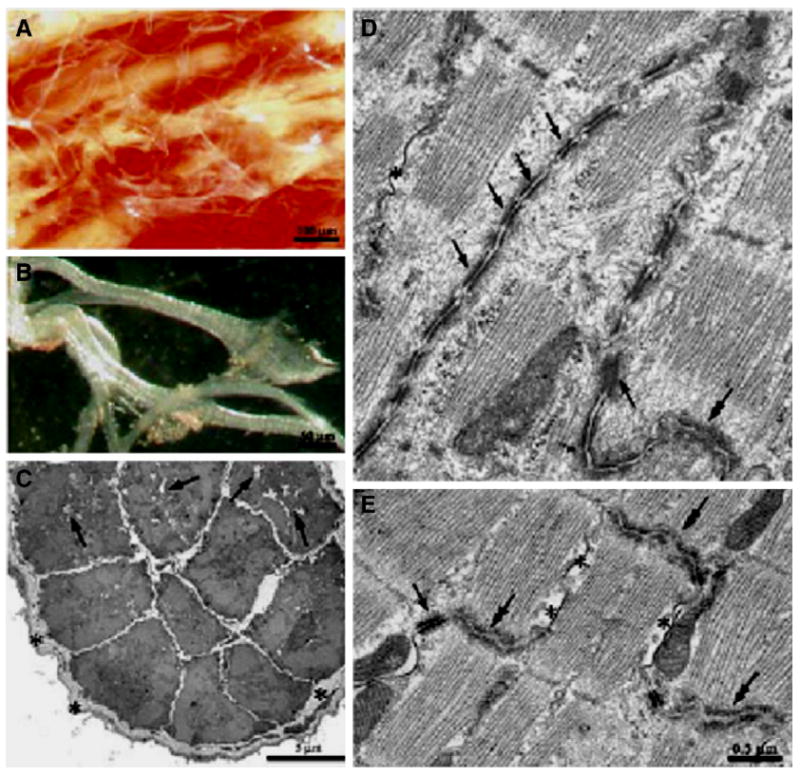
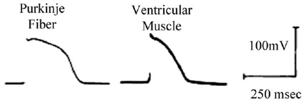
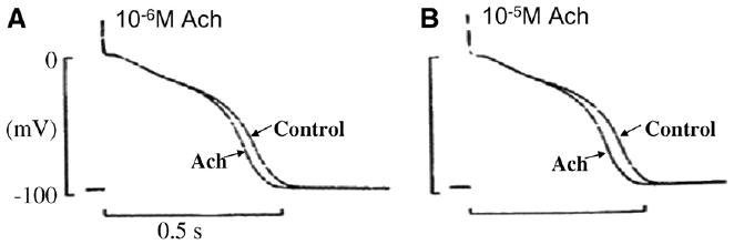
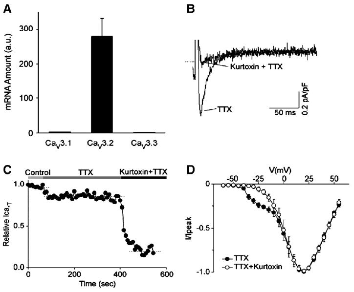
Cardiac Purkinje fibers, due to their unique anatomical location, cell structure and electrophysiologic characteristics, play an important role in cardiac conduction and arrhythmogenesis. Purkinje cell action potentials are longer than their ventricular counterpart, and display two levels of resting potential. Purkinje cells provide for rapid propagation of the cardiac impulse to ventricular cells and have pacemaker and triggered activity, which differs from ventricular cells. Additionally, a unique intracellular Ca2+ release coordination has been revealed recently for the normal Purkinje cell. However, since the isolation of single Purkinje cells is difficult, particularly in small animals, research using Purkinje cells has been restricted. This review concentrates on comparison of Purkinje and ventricular cells in the morphology of the action potential, ionic channel function and molecular determinants by summarizing our present day knowledge of Purkinje cells.


Figures

Fig. 1
Fig. 1
Panels A (low power), B (high power), Examples of Purkinje fiber strands from rat ventricle. Strands can be isolated from ventricular tissue and are generally surrounded by a thick layer of collagen. Panel C illustrates a Purkinje fiber strand that has been cut in cross section. Note collagen (asterisks) and empty cell areas filled with glycogen (arrows). Panel D illustrates very well the intercalated disk region between connecting Purkinje cells. Note the long finger like projections that form this connection. This differs substantially from the well-known staircase appearance of the disk region between two ventricular cells (Panel E). Modified from [4].
Fig. 2
Fig. 2
Fine tipped microelectrode recordings from a canine Purkinje fiber strand and ventricular muscle, paced at basic CL=500 ms and [K]o=4 mM. Note the marked differences in action potential duration (APD). Modified from [16].
Fig. 3
Fig. 3
Effects of 10−6 M (Panel A) and 10−5 M (Panel B) acetylcholine (Ach) on action potentials of a canine Purkinje fiber in normal Tyrode's solution. The stimulation rate is 1 Hz. Note that APD is dose-dependently reduced by Ach. The upper ends of the 100 mV calibration bars indicate zero potential levels. From [24].
Fig. 4
Fig. 4
Panel A, a bar graph of relative mRNA abundance for the Cav3.1, Cav3.2 and Cav3.3 genes in canine Purkinje fibers. The mRNA quantities are determined by real-time PCR and are represented in arbitrary units. Note that the Cav3.2 gene is expressed at 100-fold more abundance than the Cav3.1 and Cav3.3 genes. Panel B, Kurtoxin selectively blocks the T-type component of the whole cell calcium current in canine Purkinje cells, consistent with Cav3.2 underlying this current. The current is elicited by a test pulse at − 25 mV from a holding potential of −90 mV, in control conditions and after the application of 200 nM kurtoxin, as indicated. Both control and drug recordings are performed in the presence of 20 μM TTX. Panel C, time course for the T-type calcium current inhibition by Kurtoxin. The horizontal bars on top of the graph indicate the bath solution applied. Panel D, average current–voltage plots obtained under control conditions and after application of Kurtoxin. The holding potential is −70 mV and test pulses are applied from −60 to +55 mV at 5 mV intervals. Modified from [37].
Fig. 5
Fig. 5
Recovery of Ito from inactivation, as determined with paired 100 ms pulses (P1 and P2) delivered at 0.1 Hz from −80 to +50 mV with various P1–P2 intervals. Panel A and B, recording at P1–P2 intervals from a representative ventricular myocyte and a Purkinje cell, respectively. Panel C and D, ratios of current during P2 (I2) to current during P1 (I1) as a function of P1–P2 interval at HPs of −80 and −60 mV in canine ventricular and Purkinje cells, respectively. Best-fit biexponential functions are shown. Panel E, frequency dependence of Ito as determined by the ratio of the current during the 15th pulse to the current during the 1st pulse of a train of 100-ms pulses from −80 to +50 mV. Trains are separated by 60 s at the HP. VM: ventricular cells; PC: Purkinje cells. **P<0.01 versus ventricular cells. Note that Ito is more use dependent in Purkinje cells than ventricular cells. Modified from [44].
Fig. 6
Fig. 6
Recording of canine Purkinje cell (Panel A) and ventricular cell (Panel B) Ito on depolarization to +50 mV before (CTL) and during exposure to a series of 4-aminopyridine (4-AP) concentrations. Panel C, concentration–response curves for Ito inhibition by 4-AP, along with Hill equation fits. Panel D, Ito recording from a ventricular cell before and after exposure to 10 mM tetraethylammonium (TEA), respectively. E, Ito recording from a Purkinje cell before and after exposure to 10 mM TEA, respectively. Inset shows the Purkinje cell Ito density in absence and presence of 10 mM TEA. *P<0.05 versus control. PC: Purkinje cells; VM: Ventricular cells. Note a large TEA-sensitive outward current exists in Purkinje and not ventricular cells. Modified from [49].
Fig. 7
Fig. 7
The slow time-dependent inward currents were elicited upon hyperpolarization from a holding potential of −50 mV. Panel A: If current traces from a ventricular cell. Panel B: If current traces from a canine Purkinje cell. Panel C: Activation–voltage relation of If in the canine ventricular (○) and Purkinje (●) cells. Note that there is a significant positive shift (30 mV) of the activation curve of If in the Purkinje cell. Modified from [75].

Similar articles

Cited by

References

    1. Weerasooriya R, Hsu LF, Scavee C, Sanders P, Hocini M, Cabera JA, et al. Catheter Ablation of ventricular fibrillation in structurally normal hearts targeting the RVOT and Purkinje ectopy. Herz. 2003;28:598–606. - PubMed
    1. Lopera G, Stevenson WG, Soejima K, Maisel WH, Koplan B, Sapp JL, et al. Identification and ablation of three types of ventricular tachycardia involving the His–Purkinje system in patients with heart disease. J Cardiovasc Electr. 2004;15:52–8. - PubMed
    1. Kleber AG, Rudy Y. Basic mechanisms of cardiac impulse propagation and associated arrhythmias. Physiol Rev. 2004;84:431–88. - PubMed
    1. Di Maio A, Ter Keurs HEDJ, Franzini-Armstrong C. T-tubular profiles in Purkinje fibres of mammalian myocardium. J Musc Res Cell Motil. 2007;28:115–21. - PubMed
    1. Desplantez T, Dupont E, Severs N, Weingart R. Gap junction channels and cardiac impulse propagation. J Membr Biol. 2007;218:13–28. - PubMed

Publication types