. 2008 Sep 11;4(3):260-270.
doi: 10.1016/j.chom.2008.07.008.

Acyclovir is activated into a HIV-1 reverse transcriptase inhibitor in herpesvirus-infected human tissues

Affiliations

Acyclovir is activated into a HIV-1 reverse transcriptase inhibitor in herpesvirus-infected human tissues

Andrea Lisco et al. Cell Host Microbe. .

Abstract

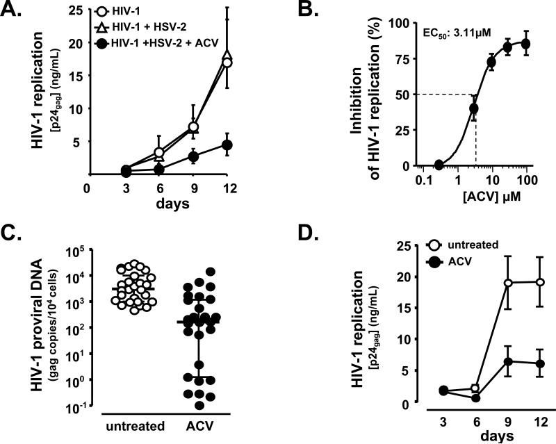
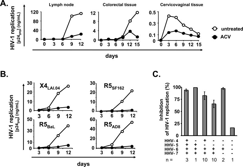
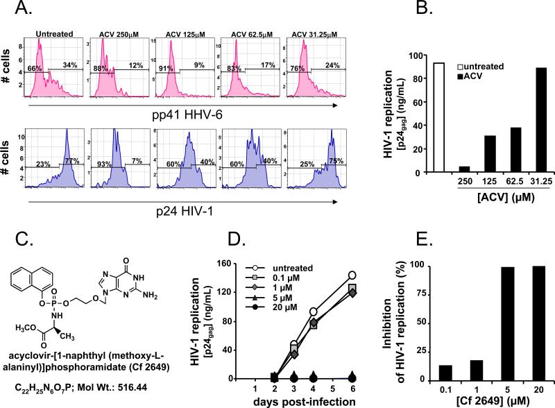
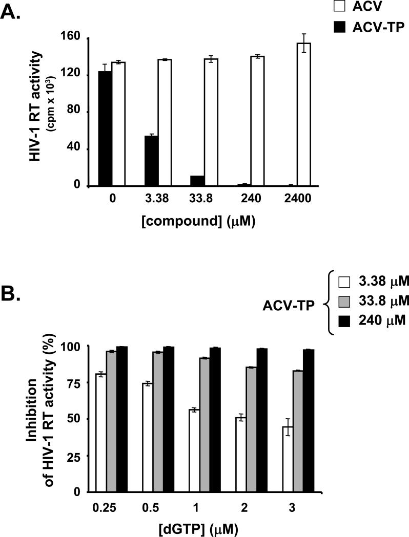
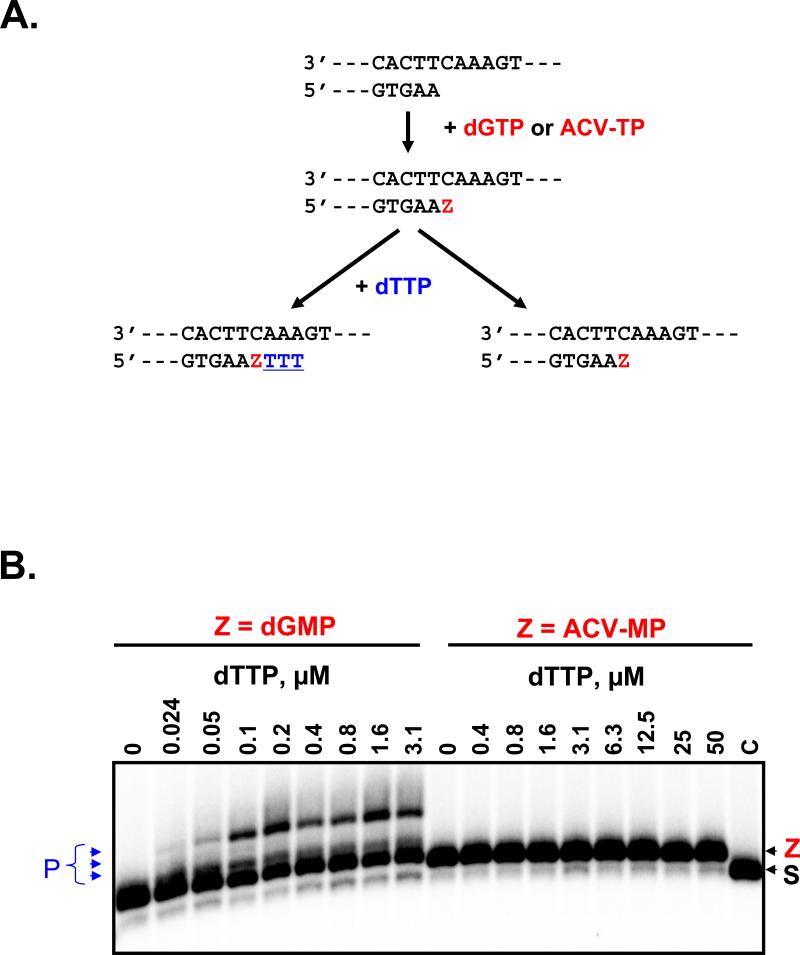
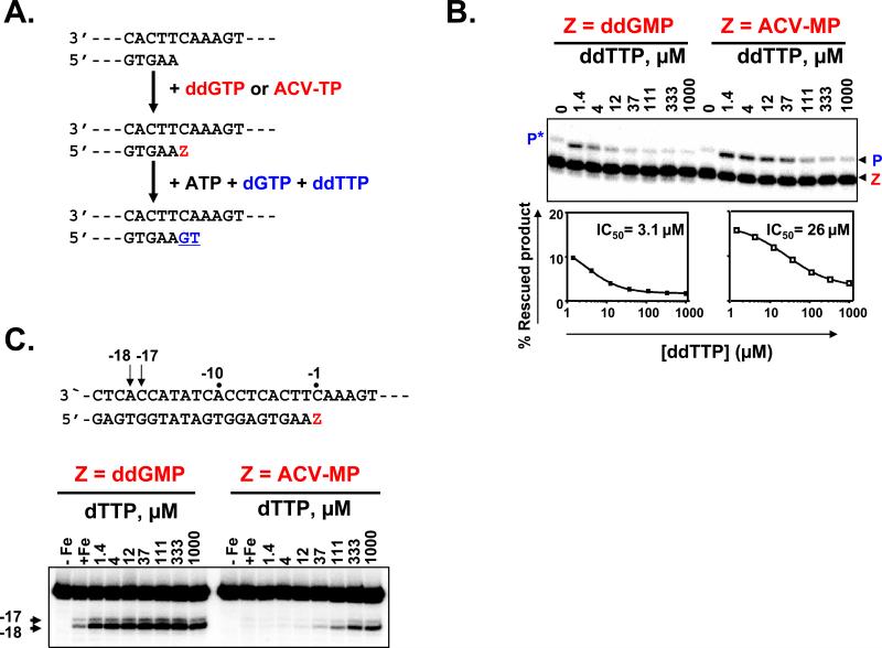
For most viruses, there is a need for antimicrobials that target unique viral molecular properties. Acyclovir (ACV) is one such drug. It is activated into a human herpesvirus (HHV) DNA polymerase inhibitor exclusively by HHV kinases and, thus, does not suppress other viruses. Here, we show that ACV suppresses HIV-1 in HHV-coinfected human tissues, but not in HHV-free tissue or cell cultures. However, addition of HHV-6-infected cells renders these cultures sensitive to anti-HIV ACV activity. We hypothesized that such HIV suppression requires ACV phosphorylation by HHV kinases. Indeed, an ACV monophosphorylated prodrug bypasses the HHV requirement for HIV suppression. Furthermore, phosphorylated ACV directly inhibits HIV-1 reverse transcriptase (RT), terminating DNA chain elongation, and can trap RT at the termination site. These data suggest that ACV anti-HIV-1 activity may contribute to the response of HIV/HHV-coinfected patients to ACV treatment and could guide strategies for the development of new HIV-1 RT inhibitors.


Figures

Figure 1
Figure 1. ACV suppresses HIV infection in human tonsillar tissues
a. Blocks of human tonsillar tissue were co-inoculated ex vivo with HSV-2 strain G and X4LAI.04, and treated or not with ACV (30 μM). HIV-1 replication was monitored by measuring p24gag accumulated in culture media over 3 day periods. Presented are means ± SEM of the results with tissues from 4 to 17 donors. For each donor, each data point represents pooled viral release from 27 tissue blocks. Note that ACV suppresses HIV-1 in HSV-2 coinfected tissues. b. HIV-1 replication was measured in tonsillar blocks infected with X4LAI.04 as in Fig.1a. ACV was added at the concentrations of 0.3, 3, 10, 30, and 100 μM, and its anti-HIV activity was evaluated from the suppression of viral replication compared with donor-matched HIV-infected tissues not treated with ACV. The 50% effective concentration (EC50) was estimated by fitting the data to 4 parameter logistic regression and was estimated to be 3.1 μM (95% confidence interval: 1.85-5.24). Presented are means ± SEM of the results with tissues from 3 to 7 donors Note that ACV suppresses HIV-1 replication in the tonsillar tissues in a dose-dependent manner. c. HIV-1 proviral DNA load in tissue blocks at day 12 post-infection was measured by real time PCR. Presented are medians and interquartile ranges of the results, (n = 27). Note that ACV efficiently reduces HIV-1 proviral DNA load in blocks of tonsillar tissue. d. HIV-1 replication was monitored as in Fig. 1a. Presented are means ± SEM of the results, (n = 38). Note that ACV efficiently suppresses HIV-1 replication in the tonsillar tissues tested.
Figure 2
Figure 2. ACV suppresses replication of different HIV-1 variants in human tissues coinfected with various HHVs
a. HIV-1 replication in blocks of human lymph nodes, colorectal and cervicovaginal tissues was monitored as in Fig.1a. For each type of tissue, the graph represents a typical result of 3 to 7 experiments performed with tissues from different donors. Note that ACV efficiently suppresses replication of HIV-1 in lymph node, colorectal and cervicovaginal tissues. b. Examples of ACV suppression of different HIV-1 variants (X4LAI.04, R5BaL, R5SF162, and R5AD8). Each data point represents pooled viral release from 27 tissue tonsillar blocks. Note that ACV efficiently suppresses replication of all four HIV-1 variants. c. Presence of HHV-1, -2, -3, -4, -5, -6, -7 and -8 by real-time PCR in blocks of tonsillar tissue. All tissues were negative for HHV-1, -2, -3, and -8. Presented are means ± SEM of the results with tonsils from n donors. Note that there are no significant differences in the level of ACV suppression of HIV-1 replication in tissues infected with various HHVs.
Figure 3
Figure 3. ACV suppresses HIV-1 in HHV-free MT-4 cell cultures either in the presence of HHV-6 infected cells or as monophosphorylated prodrug
a. HIV-1LAI.04 -infected MT4 cells were co-cultured with HHV-6B-infected MT4 cells in a ratio 10:1. Presented are the distributions of HHV-6B (upper panel) and HIV-1 LAI.04-infected cells (lower panel) as measured by flow cytometry at day 3 post-HIV-1 infection, in cultures untreated or treated with various concentrations of ACV. Note that the fraction of both HHV-6B- and HIV-1-infected cells is reduced in a dose dependent manner by ACV treatment. b. HIV-1LAI.04 -infected MT4 cells were co-cultured with HHV-6B infected MT4 cells. HIV-1 replication was monitored as in Fig.1a. Note that the replication of HIV-1 in HHV-6B/HIV-1-infected co-cultures is suppressed by ACV treatment in a dose dependent manner. c. The compound acyclovir-[1-naphthyl (methoxy-L-alaninyl)]phosphoramidate (Cf2649) has been synthesized as described in Supplemental data. d. HIV-1 LAI.04 -infected HHV-free MT4 were treated with various concentrations of the ACV monophosphorylated prodrug Cf2649. Presented are the kinetics of HIV-1LAI.04 replication, monitored by measuring p24gag accumulated in culture media at day 2, 3, 4 and 6 post-infection. Presented data are representative of two experiments. Note that compound Cf2649 suppresses the replication of HIV-1LAI.04 in a dose dependent manner. e. HIV-1LAI.04 -infected MT4 cells not infected with any HHVs were treated with various concentrations of the compound Cf2649. The cumulative release of p24gag into culture media over 6 days of culture treated with various concentrations of the compound Cf2649 is presented as fraction of the p24gag production in the untreated cultures. Presented data are representative of two independent experiments. Note that compound Cf2649 suppresses the replication of HIV-1LAI.04 in a dose-dependent manner.
Figure 4
Figure 4. ACV-TP inhibits HIV-1 RT in exogenous template reverse transcriptase assays
a. Exogenous template reverse transcriptase assays were performed in presence of various concentrations of ACV or ACV-TP as described in Experimental Procedures. Presented are means ± SEM of the results of two experiments performed in duplicates. Note that ACV-TP inhibits HIV-1 RT in a dose-dependent manner. b. The dependence of the RT inhibition by ACV-TP on the concentration of dGTP was evaluated using an exogenous template reverse transcriptase assay. dGTP was used at the indicated concentrations. The reactions were performed in the presence of the indicated concentrations of ACV-TP. Presented are means ± SEM of the results of two experiments performed in duplicate. Note that inhibition of HIV-1 RT activity by ACV-TP is inversely dependent on the concentration of dGTP.
Figure 5
Figure 5. Chain-termination with ACV-MP
a. The reaction scheme shows the relevant region of the primer and template and two possible outcomes of the primer elongation. Elongation of the primer in the presence of dGTP or ACV-TP is indicated by the “Z” in red, which refers to the incorporated monophosphate dGMP or ACV-MP, respectively. Incorporation events following position “Z” with dTTP are in blue underlined. b. The primer (S, lane C) was initially elongated to incorporate dGMP or ACV-MP, respectively at the 3’-end referred to as “Z”. Incubation with increasing concentrations of the next nucleotide dTTP resulted in three nucleotide incorporation events with the dGMP terminated primer, that are labeled as products (P). Note that the ACV-MP terminated primer is not extended, which shows that the inhibitor acts as a chain-terminator.
Figure 6
Figure 6. Inhibition of ATP-dependent excision in the presence of the next nucleotide substrate
ATP-dependent excision was monitored in a combined excision/rescue assay as previously described in Supplemental Experimental Procedures. a. The reaction scheme shows the relevant region of the primer and template. Extension of the primer in the presence of ddGTP or ACV-TP is indicated by the “Z” in red. The excision of a DNA chain-terminator requires the presence of ATP as pyrophosphate donor. The 3’-ultimate nucleotide (Z) was excised with ATP, and the simultaneous presence of dGTP and ddTTP (in blue) allowed the rescue of DNA synthesis. b. The combined excision/rescue reaction was studied with ddGMP and ACV-MP terminated primers. “Z” refers to the terminated primer and “P” to the rescued product. The asterisk indicates dGMP misincorporation in the absence of the correct ddTTP substrate. Quantification of excision/rescue reactions for ddGMP and ACV-MP are plotted. The concentration of the next complementary nucleotide required to inhibit 50% of the reaction is calculated on the basis of these two curves. Note that ACV-MP-terminated primer excision/rescue reaction is inhibited due to dead end complex formation. c. Incorporation of ACV-TP produces a dead-end complex with HIV-1 RT. Presented is site-specific footprinting of HIV-1 RT with ddGMP and ACV-MP terminated primers in the presence of increasing concentrations of the next complementary nucleotide. Lanes “–Fe” and “+Fe” show control reactions in the absence and presence of divalent Fe2+ ions that cause site-specific cleavage on the labeled template. The arrows and the sequence underneath the gel show the position of the oxidative cleavage on the template strand at positions –17 and –18, which are indicative for post- and pre-translocated complexes, respectively. Note that for ACV-MP terminated primer, at low next complementary nucleotide concentrations no cleavage occurs indicating that the complex between RT and terminated primer is fragile whereas at higher concentrations of the next complementary nucleotide RT is blocked in a dead-end complex.

Comment in

References

    1. Delta Coordinating Committee and Delta Virology Committee HIV-1 RNA response to antiretroviral treatment in 1280 participants in the Delta Trial: an extended virology study. Aids. 1999;13:57–65. - PubMed
    1. Baeten JM, S. L., Lucchetti A, Whittington WLH, Sanchez J, Coombs RW, Margaret A, Wald A, Corey L, Celum C. Herpes simplex virus suppressive treatment decreases plasma HIV-1 load in HSV-2/HIV-1 co-infected: a randomised, placebo-controlled, cross-over trial. 17th International Society fpr Sexually Transmitted Diseases Research Meeting; Seattle, WA. 2007.
    1. Bapat AR, Bodner AJ, Ting RC, Cheng YC. Identification and some properties of a unique DNA polymerase from cells infected with human B-lymphotropic virus. J Virol. 1989;63:1400–1403. - PMC - PubMed
    1. Barral K, Hider RC, Balzarini J, Neyts J, De Clercq E, Camplo M. Synthesis and antiviral evaluation of 3-hydroxy-2-methylpyridin-4-one dideoxynucleoside derivatives. Bioorg Med Chem Lett. 2003;13:4371–4374. - PubMed
    1. Burrows FJ, Gore M, Smiley WR, Kanemitsu MY, Jolly DJ, Read SB, Nicholas T, Kruse CA. Purified herpes simplex virus thymidine kinase retroviral particles: III. Characterization of bystander killing mechanisms in transfected tumor cells. Cancer Gene Ther. 2002;9:87–95. - PubMed

Publication types

MeSH terms