. 2008 Nov 7;283(45):31142-52.
doi: 10.1074/jbc.M805024200. Epub 2008 Sep 8.

An unusual intrinsically disordered protein from the model legume Lotus japonicus stabilizes proteins in vitro

Affiliations

An unusual intrinsically disordered protein from the model legume Lotus japonicus stabilizes proteins in vitro

Svend Haaning et al. J Biol Chem. .

Abstract

Intrinsic structural disorder is a prevalent feature of proteins with chaperone activity. Using a complementary set of techniques, we have structurally characterized LjIDP1 (intrinsically disordered protein 1) from the model legume Lotus japonicus, and our results provide the first structural characterization of a member of the Lea5 protein family (PF03242). Contrary to in silico predictions, we show that LjIDP1 is intrinsically disordered and probably exists as an ensemble of conformations with limited residual beta-sheet, turn/loop, and polyproline II secondary structure. Furthermore, we show that LjIDP1 has an inherent propensity to undergo a large conformational shift, adopting a largely alpha-helical structure when it is dehydrated and in the presence of different detergents and alcohols. This is consistent with an overrepresentation of order-promoting residues in LjIDP1 compared with the average of intrinsically disordered proteins. In line with functioning as a chaperone, we show that LjIDP1 effectively prevents inactivation of two model enzymes under conditions that promote protein misfolding and aggregation. The LjIdp1 gene is expressed in all L. japonicus tissues tested. A higher expression level was found in the root tip proximal zone, in roots inoculated with compatible endosymbiotic M. loti, and in functional nitrogen-fixing root nodules. We suggest that the ability of LjIDP1 to prevent protein misfolding and aggregation may play a significant role in tissues, such as symbiotic root nodules, which are characterized by high metabolic activity.


Figures
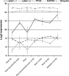
FIGURE 1.
FIGURE 1.
Steady state level of LjIdp1 mRNA determined by microarray analysis. Expression of LjIdp1 in different root tissues, inoculated roots, and mature nodules. LjIdp1 is represented on the chip with two probe sets (LjIdp1_1 and LjIdp1_2) with different efficiencies. Legend code example, “Root 1d Rz” is roots 1 day postinoculation with Rhizobium. PP2A, protein phosphatase 2A; GAPDH, glyceraldehyde-3-phosphate dehydrogenase.
FIGURE 2.
FIGURE 2.
Relative expression level of LjIdp1 in different plant organs and roots 2 days post inoculation with Rhizobium as determined by real time PCR. Error bars, S.D. values.
FIGURE 3.
FIGURE 3.
SR-CD spectroscopy of LjIDP1. A, the SR-CD spectra of LjIDP1 showing the effect of temperature on the secondary structure. The spectrum of LjIDP1 has a large negative peak at ∼200 nm typical for an unfolded polypeptide and a small negative ellipticity at ∼220 nm due to β-sheet. Heating the sample from 20 to 80 °C has little effect on the CD spectrum. Note the small intensity and shape of the difference spectrum Δ(20-80 °C) that is similar to the CD spectrum of PPII. B, 70 consecutive SR-CD scans of LjIDP1 undergoing dehydration were collected. Virtually no changes were observed during the first 18 scans, so these were averaged and denoted “wet state.” All major structural changes occurred after 39 scans; therefore, spectra 40-70 were averaged and denoted “dry state.” The spectrum of LjIDP1 in the dry state is characterized by a positive peak at ∼190 nm and negative ellipticity at ∼208 nm and ∼222 nm typical for α-helix. C, SR-CD spectroscopy of LjIDP1 in the presence of different detergents and alcohols. Intriguingly, all agents tested induced α-helix at the expense of the β-sheet and disordered fractions. D, the table shows the quantified relative amounts of secondary structure elements.
FIGURE 4.
FIGURE 4.
Aliphatic/methyl region of one-dimensional NMR spectra of LjIDP1 in buffer (a) and in buffer plus 6 m urea (b). The low dispersion in a indicates that LjIDP1 lacks well defined structure, and the high similarity between the two spectra indicates that no significant structural rearrangements have occurred.
FIGURE 5.
FIGURE 5.
Size exclusion chromatography suggests that LjIDP1 belongs to the native premolten globule class of IDPs. The Stokes radius (Rs) of LjIDP1 was determined by gel filtration chromatography on a Superdex 75 HR 10/300 GL column. LjIDP1 elutes at 11.3 ml, which corresponds to a Stokes radius of 24.5 Å. The empirical equations (trend lines) representing the dependences of the hydrodynamic radii (Rs) on the molecular mass for different protein conformational states is from Uversky (16).
FIGURE 6.
FIGURE 6.
Protease sensitivity assay. LjIDP1, α-synuclein, and lysozyme were incubated with decreasing concentrations of trypsin for 1 h at 37 °C and analyzed by SDS-PAGE on a 10-20% polyacrylamide gradient gel. The numbers on the gels represent the relative fraction of trypsin (w/w).
FIGURE 7.
FIGURE 7.
FTIR spectrum of dehydrated LjIDP1. Curve fitting with five Lorentzian peaks produced an excellent fit. Curve 3 (1657 cm-1) was assigned to α-helix, curve 2 (1634 cm-1) to β-sheet, and curve 4 (1681 cm-1) to turns.
FIGURE 8.
FIGURE 8.
Composition profiling of LjIDP1 and homologous proteins (28IDP1). A, plot of mean hydrophobicity (H) against mean net charge (R) of 28IDP1 (LjIDP1 (gray circle) and 27 homologous proteins). This group has very similar R and H values, and interestingly, all are located below the boundary line separating small globular proteins (below the line) from IDPs (above the line). The boundary line satisfies the equation, H = (R + 1.157)/2.785. B, amino acid composition profiles of Disprot 3.4 versus Swissprot (first table) and 28IDP1 versus Disprot 3.4 (second table). Residues overrepresented in Disprot 3.4 compared with Swissprot 51 are defined as disorder-promoting, and residues underrepresented are defined as order-promoting. It is evident that disorder-promoting residues are mainly hydrophilic, whereas order-promoting residues are mainly hydrophobic. The subsequent profiling of 28IDP1 versus Disprot 3.4 reveals that all except five residues occur with significantly different frequencies in these data sets and that 28IDP1 has less disorder-promoting and more order-promoting residues than DisProt 3.4.
FIGURE 9.
FIGURE 9.
The cumulative distribution function curve of LjIDP1 PONDR VL-XT scores superimposes on the boundary line separating ordered proteins (above) from disordered proteins (below).
FIGURE 10.
FIGURE 10.
LjIDP1 prevents dehydration- and freezing-induced enzyme inactivation. A, residual activity of CS (250 μg/ml) before and after repeated cycles of dehydration/rehydration either alone of in the presence of stabilizing agents. C, similar experiment with LDH (100 μg/ml). LjIDP1 effectively prevents inactivation of CS and LDH even after four cycles of dehydration/rehydration. B, residual activity of CS (250 μg/ml) before and after repeated cycles of freezing/thawing, either alone or in the presence of stabilizing agents. D, similar experiment with LDH 100 μg/ml. CS alone is only slightly affected by freezing, and after eight cycles of freezing/thawing, it retains ∼60% activity. In contrast, LDH alone quickly loses activity upon freezing, but adding LjIDP1 effectively prevents this. Numbers on the x axis refer to the number of cycles. The activity at the start (cycle 0) of the experiments is set to 100%. Error bars, 95% confidence intervals.

Similar articles

Cited by

References

    1. Uversky, V. N., Oldfield, C. J., and Dunker, A. K. (2008) Annu. Rev. Biophys. 37 215-246 - PubMed
    1. Tunnacliffe, A., and Wise, M. J. (2007) Naturwissenschaften 94 791-812 - PubMed
    1. Dyson, H. J., and Wright, P. E. (2005) Nat. Rev. Mol. Cell Biol. 6 197-208 - PubMed
    1. Colebatch, G., Desbrosses, G., Ott, T., Krusell, L., Montanari, O., Kloska, S., Kopka, J., and Udvardi, M. K. (2004) Plant J. 39 487-512 - PubMed
    1. Walsh, K. B., Vessey, J. K., and Layzell, D. B. (1987) Plant Physiol. 85 137-144 - PMC - PubMed

Publication types

MeSH terms

Substances