. 2008 Nov;76(11):5082-92.
doi: 10.1128/IAI.00698-08. Epub 2008 Sep 8.

Characterization of a unique ClpB protein of Mycoplasma pneumoniae and its impact on growth

Affiliations

Characterization of a unique ClpB protein of Mycoplasma pneumoniae and its impact on growth

T R Kannan et al. Infect Immun. 2008 Nov.

Abstract

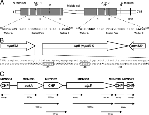
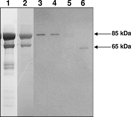
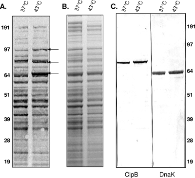
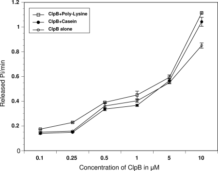
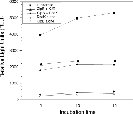
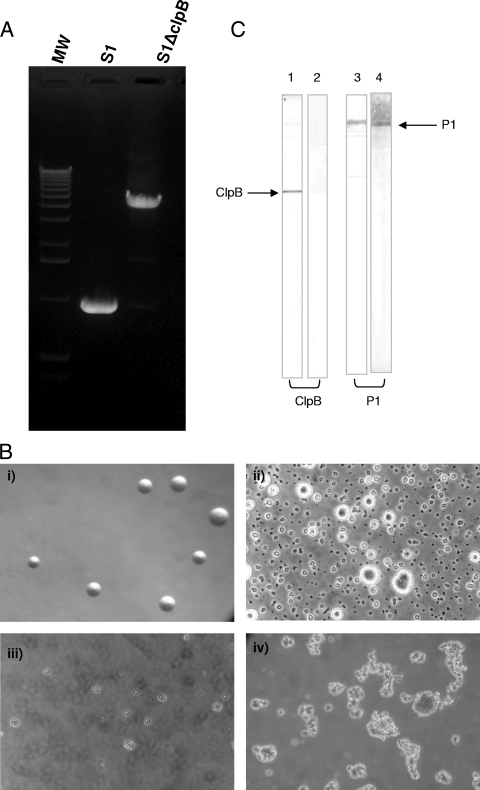
Mycoplasma pneumoniae accounts for 20 to 30% of all community-acquired pneumonia and has been associated with other airway pathologies, including asthma, and a range of extrapulmonary manifestations. Although the entire genomic sequence of M. pneumoniae has been completed, the functions of many of these genes in mycoplasma physiology are unknown. In this study, we focused on clpB, a well-known heat shock gene in other bacteria, to examine its role in mycoplasma growth. Transcriptional and translational analyses of heat shock in M. pneumoniae indicated that clpB is significantly upregulated, reinforcing its status as a critical responder to heat stress. Interestingly, M. pneumoniae ClpB does not use dual translational start points for ClpB synthesis, like other ClpB-characterized bacteria. Biochemical characterization of purified M. pneumoniae recombinant ClpB revealed casein- and lysine-independent ATPase activity and DnaK-DnaJ-GrpE-dependent chaperone activity. An M. pneumoniae mini-Tn4001-integrated, clpB-null mutant was impaired in its ability to replicate under permissive growth conditions, demonstrating the growth-promoting status of ClpB.


Figures

FIG. 1.
FIG. 1.
M. pneumoniae ClpB organization and transcription. (A) Proposed domain organization of M. pneumoniae ClpB and conserved motifs involved in ATP binding and ATP hydrolysis. M. pneumoniae ClpB retains only a shortened N-terminal domain, two nucleotide binding domains (ATP-1, positions 115 to 265; and ATP-2, positions 392 to 583) which are separated by the middle region, and a C-terminal region. Both nucleotide binding domains contain the Walker A (60GX4GKT69 and 466GX4GKT473) and Walker B (130Hy2DE133 and 536Hy2DE539) motifs, where Xn represents n amino acids of any kind and Hyn represents n hydrophobic amino acids. ATP-1 has one additional nucleotide binding motif, termed Walker B′ (249Hy2DE252). CPs (positions 102 to 111 and 511 to 519) contribute to protein substrate binding. Arg (R) residues (positions 187 and 609) are proposed to serve as Arg fingers, contacting ATP bound to an adjacent subunit. The sensor and substrate domain (SSD) (666GAR668) potentially senses the nucleotide status of ATP-2. The middle coiled-coil region is predicted to be involved in protein-protein interactions. (B) Promoter region of clpB. The bold ATG (far right) indicates the putative translational start site of clpB (mpn531). Inverted repeats on each side of the 9-nucleotide spacer sequence of the CIRCE element are shown in bold. The promoter region contains a Pribnow box (−10 site), which is an exact match for the consensus sequence (TATAAT) for the housekeeping sigma factor (σ70). A putative −35 site (boxed) was also identified upstream of the Pribnow box. The stop codon of mpn532 (TAG, far left) is 175 nucleotides upstream of the start codon of clpB. An asterisk designates the transcriptional start site (bold letter) as determined by PE. A Shine-Dalgarno (SD) sequence (underlined) is located downstream of the putative transcriptional start site. (C) Transcriptional analysis of genes surrounding clpB of M. pneumoniae. RT-PCR was performed on total RNA isolated from M. pneumoniae cells as described in Materials and Methods. A schematic of clpB and its surrounding genes is presented. Conserved hypothetical proteins (CHP) are indicated. Thin arrow lines represent regions that were amplified to confirm transcription of individual genes. Bold arrow lines represent RT-PCR products to indicate cotranscription of corresponding genes. Numbers represent the sizes of RT-PCR-amplified products.
FIG. 2.
FIG. 2.
Purification of ClpB of M. pneumoniae. rClpB protein was purified through different columns as described in Materials and Methods, separated on SDS-PAGE gel, and Coomassie blue stained. Protein fractions were from a Ni-nitrilotriacetic acid column (lane 1), a Mono Q anion exchange column (lane 2), and a Superdex 200 gel filtration column eluted using a linear NaCl gradient (lanes 3 to 6).
FIG. 3.
FIG. 3.
Translational response of M. pneumoniae during heat shock. [35S]methionine-cysteine was added to M. pneumoniae S1 cells grown in DMEM at 37°C and 43°C, and levels of radiolabeled proteins were analyzed after heat shock as described in Materials and Methods. M. pneumoniae proteins were separated on 4 to 12% Nu-PAGE gel and transferred to nitrocellulose membranes and exposed to X-ray film (A), Coomassie blue stained (B), and transferred to nitrocellulose membranes and immunoblotted using anti-ClpB and anti-DnaK antisera (C). Arrows indicate the upregulated heat shock proteins.
FIG. 4.
FIG. 4.
ATPase activities of ClpB. ATPase activities of ClpB were measured at different ClpB protein concentrations as described in Materials and Methods in the absence or presence of α-casein (0.25 mg/ml) or poly-l-lysine (0.04 mg/ml).
FIG. 5.
FIG. 5.
Chaperone activities of ClpB. The activities of firefly luciferase during its chaperone-assisted reactivation were monitored using luminescence (see Materials and Methods). Activities of rClpB alone; rClpB with DnaK; rClpB with DnaK, DnaJ, and GrpE (KJE); DnaK alone; and unheated luciferase alone are compared. The protein concentrations used were 33 nM luciferase, 2.0 μM ClpB, 2.0 μM M. genitalium rDnaK, 1.0 μM E. coli DnaK, 1.0 μM DnaJ, and 1.0 μM GrpE.
FIG. 6.
FIG. 6.
Construction of ClpB-null mutant M. pneumoniae S1ΔClpB. (A) PCR amplification of clpB in ClpB mutant and wild-type M. pneumoniae S1. After sequence confirmation of integration of mini-Tn4001 in clpB, the integrated region was amplified by region-specific mpn531 primers. Due to integration of Tn, an extra DNA fragment, 3 kb larger than that in wild-type M. pneumoniae S1, was amplified in mutant S1ΔClpB. MW, molecular weight. (B) Morphology of ClpB mutant of M. pneumoniae S1 strain. (i) ClpB mutant on SP-4 agar plate (similar to wild-type result [not shown]). (ii to iv) Morphologies of wild-type and ClpB mutant cells grown in SP-4 broth: (ii) M. pneumoniae S1 after 48 h (showing well-established adhering cells/colonies), (iii) ClpB mutant after 1 week (showing mostly floating cells/colonies), and (iv) ClpB mutant after 3 weeks (showing large, floating clumps). (C) ClpB expression in M. pneumoniae. Total cell lysates from wild-type M. pneumoniae strain S1 and the ClpB mutant (S1ΔClpB) were separated on SDS-PAGE gel, transferred to a nitrocellulose membrane, and treated with anti-ClpB and anti-P1 adhesin antibodies. Lanes: 1, S1 anti-ClpB antibodies; 2, S1ΔClpB anti-ClpB antibodies; 3, S1 anti-P1 antibodies; 4, S1ΔClpB anti-P1 antibodies.

References

    1. Barnett, M. E., A. Zolkiewska, and M. Zolkiewski. 2000. Structure and activity of ClpB from Escherichia coli. Role of the amino- and carboxyl-terminal domains. J. Biol. Chem. 27537565-37571. - PMC - PubMed
    1. Baseman, J. B. 1993. The cytadhesins of Mycoplasma pneumoniae and M. genitalium. Subcell. Biochem. 20243-259. - PubMed
    1. Baseman, J. B., and J. G. Tully. 1997. Mycoplasmas: sophisticated, reemerging, and burdened by their notoriety. Emerg. Infect. Dis. 321-32. - PMC - PubMed
    1. Beinker, P., S. Schlee, Y. Groemping, R. Seidel, and J. Reinstein. 2002. The N terminus of ClpB from Thermus thermophilus is not essential for the chaperone activity. J. Biol. Chem. 27747160-47166. - PubMed
    1. Benders, G. A., B. C. Powell, and C. A. Hutchison III. 2005. Transcriptional analysis of the conserved ftsZ gene cluster in Mycoplasma genitalium and Mycoplasma pneumoniae. J. Bacteriol. 1874542-4551. - PMC - PubMed

Publication types

LinkOut - more resources