. 2008 Nov;76(11):5228-37.
doi: 10.1128/IAI.00410-08. Epub 2008 Sep 8.

Outer surface protein A protects Lyme disease spirochetes from acquired host immunity in the tick vector

Affiliations

Outer surface protein A protects Lyme disease spirochetes from acquired host immunity in the tick vector

James M Battisti et al. Infect Immun. 2008 Nov.

Abstract

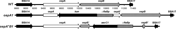
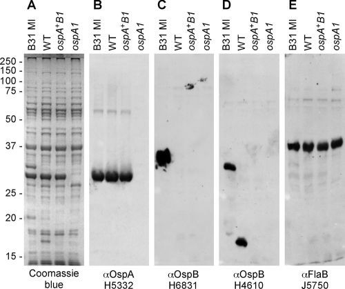
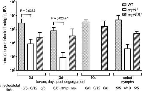
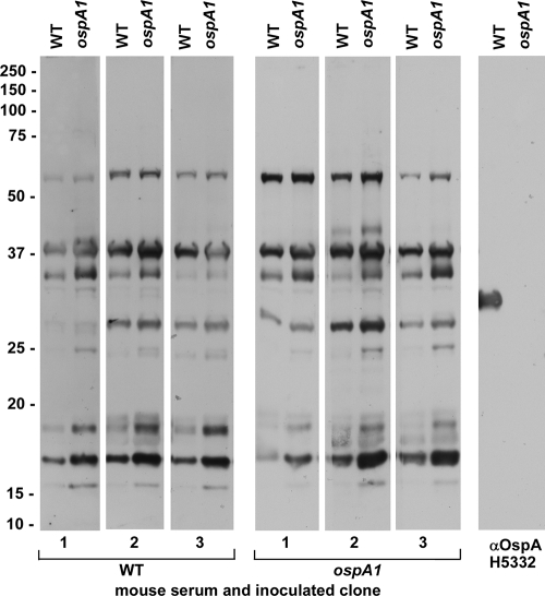
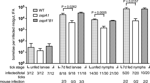
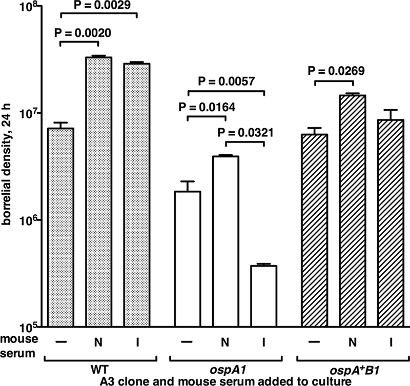
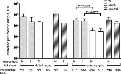
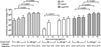
The Lyme disease spirochete Borrelia burgdorferi alters the expression of outer surface protein (osp) genes as the bacterium cycles between ticks and mammals. OspA is produced as borreliae enter the tick vector and remains a major surface antigen during midgut colonization. To elucidate the role of OspA in the vector, we created an insertional deletion of ospA in strain B31-A3. The ospA mutant infects mice when it is injected intradermally and is acquired by larval ticks fed on these mice, where it persists through the molt to the nymph stage. Bacterial survival rates in artificially infected tick larvae fed on naïve mice were compared with those in the vector fed on immune mice. The ospA mutant proliferates in larvae if it is exposed to blood from naïve mice, but it declines in density after larval feeding if the blood is from immune mice. When uninfected larvae are fed on B-cell-deficient mice infected with the ospA mutant, larvae show borrelial densities and persistence that are significantly greater than those fed on infected, immunocompetent mice. We conclude that OspA serves a critical antibody-shielding role during vector blood meal uptake from immune hosts and is not required for persistence in the tick vector.


Figures

FIG. 1.
FIG. 1.
Physical maps of the lp54 ospAB locus in the B31-A3 WT, the ospA mutant, and the ospA restored strains. WT, segment of lp54 from bp 8750 to 11400, as indicated by numbers below the map (27), from locus tag BBA14 to BBA17 in clonal isolate A3-6. ospA1, mutation of ospA by insertion of a flaBP-kan marker (10) inserted in an internally deleted ospA gene via transformation of A3-6 with pOKOSPAKO (described in Materials and Methods), resulting in clone 2E6. ospA+B1, restoration of the ospA mutation by insertion of a flaBP-aacC1 marker (22) in an internally deleted ospB gene on the 3′ end of an intact ospA gene via transformation of 2E6 with pOKOSPA-C′ (described in Materials and Methods), resulting in clone 7A.
FIG. 2.
FIG. 2.
Protein profiles of strain B31 MI and the WT and isogenic ospAB clones; detection of OspA, OspB, and FlaB. (A) Coomassie blue gel shows whole-cell lysates from 35°C cultures of B31 MI, WT, ospA+B1, and ospA1 strains separated by SDS-PAGE and stained with Coomassie blue. (B to E) Immunoblots of replicate SDS-PAGE separations reacted as described in Materials and Methods with OspA-specific monoclonal antibody H5332 (B), OspB-specific monoclonal antibody H6831 (C), OspB-specific monoclonal antibody H4610 (D), and FlaB-specific rabbit polyclonal antiserum J5750 (E). Migration of molecular mass standards in kDa is indicated at left. αOspA, anti-OspA.
FIG. 3.
FIG. 3.
Acquisition of the WT and ospAB clones from infected mice. RML mice were injected intradermally with 5 × 10³ bacteria of the WT, ospA1, and ospA+B1 clones, as designated, and normal I. scapularis larvae were allowed to feed on seropositive mice at 3 weeks postinjection. Borreliae in dissected midguts from each group were enumerated by IFA at 0, 3, and 10 days postengorgement and after they molted to nymphs (unfed nymphs); values represent means plus standard deviations (error bars) for infected midguts. Infection frequency (no. of infected/total ticks) of assayed tick groups is shown. The ospA1 and ospA+B1 groups were subjected to a t test comparison with the WT group at each time point; a significant t test P value is shown above bracket. *, the value for the WT and ospA1 clones at 3 days postengorgement was significant (P = 0.0247) compared by an unpaired t test with Welch's correction.
FIG. 4.
FIG. 4.
Recognition of B. burgdorferi antigens by sera from infected mice. RML mice were injected intradermally with 5 × 10⁴ bacteria of the WT or ospA1 clone, as designated, and serum was recovered from blood collected 8 weeks postinjection. These infected mouse sera (1:200 dilution) and OspA-specific monoclonal antibody H5332 (1:500 dilution) were reacted with immunoblots of the WT and ospA1 clones as described in Materials and Methods. Migration of molecular mass standards in kDa is indicated at left. αOspA, anti-OspA.
FIG. 5.
FIG. 5.
Replication of the WT and ospAB borreliae in artificially infected larvae fed on naïve mice. I. scapularis larvae infected by immersion in the WT, ospA1, and ospA+B1 clones as indicated were fed on RML mice at larval and nymphal stages. Borrelial density in infected midguts from larvae and nymphs before feeding or at 7 days postengorgement was enumerated by IFA; values represent means plus standard deviations (error bars) for infected midguts. The infection frequency of assayed tick groups is shown as in Fig. 3. The transmission frequency of borreliae to mice (mouse infection) was determined as described in Materials and Methods. Ticks infected with the ospA1 and ospA+B1 clones were subjected to t test comparison with the WT at each time point; significant t test P values are shown above the brackets.
FIG. 6.
FIG. 6.
Survival of the OspA-deficient and control clones with mouse serum in vitro. The WT, ospA1, and ospA+B1 clones grown in BSK II (−) or BSK II containing naïve (N) or borrelia-infected (I) mouse sera, as described in Materials and Methods. Values are mean borrelial densities plus standard deviations (error bars) per ml, from colony counts of dilutions after 24 h of incubation (duplicate incubations for each condition). One-way analysis of variance posttest values for significant differences between conditions are shown above the brackets.
FIG. 7.
FIG. 7.
Effect of acquired immunity to B. burgdorferi on the survival of OspA-deficient borreliae. Naïve, mock-infected RML mice (N) and immune mice (I) infected via larval tick bite with the A3-6 wild type, identified as seropositive and cured by ceftriaxone treatment, were used as hosts for feeding by larvae artificially infected with the WT, ospA1, and ospA+B1 clones. Borrelial densities in infected midguts from larvae 4 days postengorgement and in molted, unfed nymphs were enumerated by IFA; values represent means plus standard deviations (error bars) for infected midguts. The infection frequencies of assayed tick groups are shown as in Fig. 3. Student's t test values for significant differences between naïve- and immune-fed ticks at each tick stage are shown above the brackets.
FIG. 8.
FIG. 8.
Relative survival of OspA-deficient borreliae acquired from intact and B-cell-deficient infected mice. C57BL/6J (B6) and C57BL/6J Igh−/− (B6.Igh−/−) congenic mice were injected with the WT, ospA1, and ospA+B1 clones, and B6 mice were tested at 3 weeks for serological response; normal I. scapularis larvae were fed at 4 to 5 weeks postinfection. Borrelial density in infected midguts from larvae at 0, 3, and 10 days postengorgement was enumerated by IFA; values represent means plus standard deviations (error bars) for infected midguts. The infection frequencies of assayed tick groups are shown as in Fig. 3. Statistically significant t test values of borrelial midgut density for ticks fed on B6 mice relative to ticks fed on the B6.Igh−/− mouse host genotype at each time point are shown above the brackets; *, the value for the ospA1 clone at day 0 postengorgement, with B6 versus B6.Igh−/− mice, was significant (P = 0.0017) compared by an unpaired t test with Welch's correction.

References

    1. Anderson, J. F., R. C. Johnson, L. A. Magnarelli, and F. W. Hyde. 1985. Identification of endemic foci of Lyme disease: isolation of Borrelia burgdorferi from feral rodents and ticks (Dermacentor variabilis). J. Clin. Microbiol. 2236-38. - PMC - PubMed
    1. Barbour, A. G. 1984. Isolation and cultivation of Lyme disease spirochetes. Yale J. Biol. Med. 57521-525. - PMC - PubMed
    1. Barbour, A. G., and C. F. Garon. 1987. Linear plasmids of the bacterium Borrelia burgdorferi have covalently closed ends. Science 237409-411. - PubMed
    1. Barbour, A. G., S. L. Tessier, and S. F. Hayes. 1984. Variation in a major surface protein of Lyme disease spirochetes. Infect. Immun. 4594-100. - PMC - PubMed
    1. Barbour, A. G., S. L. Tessier, and W. J. Todd. 1983. Lyme disease spirochetes and ixodid tick spirochetes share a common surface antigenic determinant defined by a monoclonal antibody. Infect. Immun. 41795-804. - PMC - PubMed

Publication types

MeSH terms