. 2008 Sep 29;205(10):2359-68.
doi: 10.1084/jem.20080353. Epub 2008 Sep 8.

Delayed protection by ESAT-6-specific effector CD4+ T cells after airborne M. tuberculosis infection

Affiliations

Delayed protection by ESAT-6-specific effector CD4+ T cells after airborne M. tuberculosis infection

Alena M Gallegos et al. J Exp Med. .

Abstract

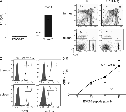
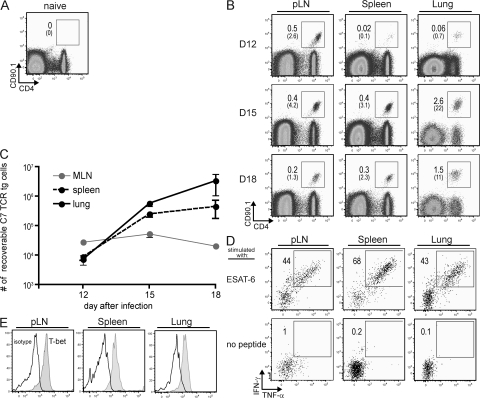
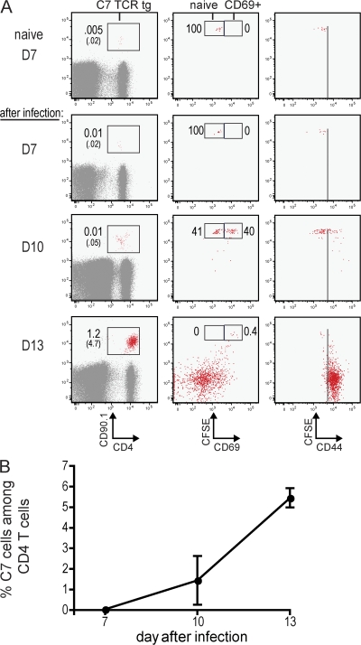
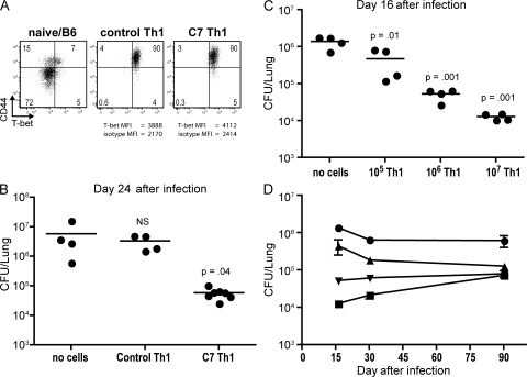
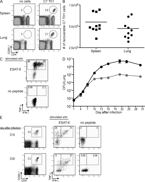
Mycobacterium tuberculosis infection induces complex CD4 T cell responses that include T helper type 1 (Th1) cells and regulatory T cells. Although Th1 cells control infection, they are unable to fully eliminate M. tuberculosis, suggesting that Th1-mediated immunity is restrained from its full sterilizing potential. Investigation into T cell-mediated defense is hindered by difficulties in expanding M. tuberculosis-specific T cells. To circumvent this problem, we cloned CD4(+) T cells from M. tuberculosis-infected B6 mice and generated transgenic mice expressing a T cell receptor specific for the immunodominant antigen early secreted antigenic target 6 (ESAT-6). Adoptively transferred naive ESAT-6-specific CD4(+) T cells are activated in pulmonary lymph nodes between 7 and 10 d after aerosol infection and undergo robust expansion before trafficking to the lung. Adoptive transfer of activated ESAT-6-specific Th1 cells into naive recipients before aerosol M. tuberculosis infection dramatically enhances resistance, resulting in 100-fold fewer bacteria in infected lungs. However, despite large numbers of Th1 cells in the lungs of mice at the time of M. tuberculosis challenge, protection was not manifested until after 7 d following infection. Our results demonstrate that pathogen-specific Th1 cells can provide protection against inhaled M. tuberculosis, but only after the first week of infection.


Figures

Figure 1.
Figure 1.
C7 TCR tg CD4+ T cells are specific to ESAT-6. (A) Clone 7 hybridoma cells or hybridoma fusion partner BW5147 cells were stimulated with 5 μg/ml of ESAT-6 peptide, or with media in the presence of APCs. Supernatants were collected 2 d later, and IL-2 levels were measured by ELISA. nd, not detectable. (B) Splenocytes and thymocytes from B6 or C7 TCR tg mice were stained for CD4 and CD8 expression. Numbers indicate the percentages of cells in each gate. (C) Vβ10b and Vα11.1/.2 expression after gating on CD4+ thymocytes or CD4+ splenocytes from the indicated mice. Shaded gray histograms represent CD4+ T cells from B6 mice; continuous lines represents CD4+ T cells from C7 TCR tg mice. Black and small gray numbers indicate the percentages of stained cells among CD4+ T cells from C7 TCR tg or B6 mice, respectively. (D) Purified CD4+ T cells from C7 TCR tg or B6 mice were stimulated with ESAT-6 peptide in the presence of APCs. Proliferation was measured by [3H]thymidine incorporation between 48 and 72 h of culture. Error bars in A and D represent SDs.
Figure 2.
Figure 2.
C7 TCR tg CD4+ T cells respond to M. tuberculosis infection in vivo. (A) 104 C7 TCR tg.RAG−/− CD4+ T cells (CD90.1) were transferred into uninfected congenically marked B6 recipients (CD90.2), and splenocytes were stained for CD4 and CD90.1 expression 7 d later. The gate marks donor-derived C7 TCR tg.RAG−/− CD4+ T cells (CD90.1+) cells, and the percentages of these cells within total cells (top) or among CD4+ T cells (bottom) are shown. (B–E) As in A, except mice were aerosol infected 1 d after T cell transfer with 100 CFU M. tuberculosis. pLNs, spleen, and lung were examined 12, 15, and 18 d later. (C) Graphic representation of results from three to four mice per time point. Error bars represent SDs. (D) C7 TCR tg.RAG−/− CD4+ T cells from the indicated organs were harvested from day 15–infected mice, and their ability to make IFN-γ and TNF-α was determined after stimulation with ESAT-6 peptide or medium alone. (E) Intracellular T-bet (shaded histogram) or isotype control staining (continuous line) of C7 TCR tg.RAG−/− CD4+ T cells taken from the indicated organs 15 d after infection. The data presented in this figure are representative of two experiments with three to four mice per group.
Figure 3.
Figure 3.
Naive C7 TCR tg CD4+ T cells are activated in the pLNs between days 7 and 10 after infection. (A) 105 CFSE-labeled C7 TCR tg.RAG−/− CD4+ T cells (CD90.1) were transferred into congenically marked B6 recipients (CD90.2) that were left uninfected (top row, naive) or aerosol infected with 100 CFU M. tuberculosis 1 d later (bottom three rows, after infection). The first column shows CD4 and CD90.1 staining of pLN cells. Gate marks donor-derived C7 TCR tg.RAG−/− CD4+ T cells (CD90.1+), and the percentage of these cells within total pLNs (top) or among CD4+ T cells (bottom) is shown. The second and third columns show CFSE intensity and CD69 staining or CD44 staining, respectively, on gated C7 TCR tg.RAG−/− CD4+ T cells. Numbers indicate the percentages of cells in the indicated gates. (B) Graphic representation of results from three to four mice per time point for the experiment described in A. Error bars represent SDs. The data presented in this figure are representative of three experiments of similar design with two to four mice per group.
Figure 4.
Figure 4.
Adoptive transfer of C7 Th1 T cells confers protection to M. tuberculosis. (A) B6 or C7 CD4+ T cells were stimulated in vitro with anti-CD3/CD28 (control Th1 cells) or ESAT-6 peptide (C7 Th1 cells), respectively, in the presence of IFN-γ and anti–IL-4 for 4 d, and were analyzed for CD44 and T-bet expression. CD4+ T cells from naive B6 spleens are shown as a staining control. Numbers represent the percentages of cells in each quadrant. MFI, mean fluorescence intensity. (B) 107 Th1-differentiated B6 or C7 CD4+ T cells were transferred into B6 recipient mice 1 d before aerosol infection with 100 CFU M. tuberculosis. 24 d later, the number of bacteria in the lungs was determined. Each symbol represents data from one mouse, and p-values compare the CFU from mice that received no cells compared with mice that received either B6 or C7 Th1 cells. The experiment was performed two times with similar results. Horizontal bars represent the mean. (C and D) Similar experiment as in B, except 107, 106, or 105 C7 Th1 cells or no cells were transferred and CFU were determined 16, 30, and 90 d after infection. This experiment was preformed one time with four mice per group. In C, each circle represents data from one mouse, and p-values compare the CFU from mice that received no cells. In D, each symbol represents four mice per group (square, 107 cells; inverted triangle, 106 cells; triangle, 105 cells; and circle, no cells). Error bars represent SDs.
Figure 5.
Figure 5.
C7 Th1 cells confer protection to M. tuberculosis only after day 7 following infection. (A–C) 107 C7 Th1 cells (CD90.1) were transferred into B6 recipient mice (CD90.2), and 1 d later splenocytes or lung cells were harvested and stained for CD4 and CD90.1 expression. (A) Gate marks donor-derived cells among total cells in the indicated organs. (B) Graphic representation showing the number of recoverable C7 Th1 cells from the indicated organs. Each symbol represents data from one mouse. (C) Intracellular TNF-α and IFN-γ staining of C7 Th1 cells harvested from the lungs. bars in B and C represent the mean. (D) Similar experiment as in A–C, except mice were infected 1 d after T cell transfer with 100 CFU M. tuberculosis. Bacterial numbers were determined at the indicated days after infection. The data presented is a compilation of five experiments. Each time point includes CFU data from 7–12 mice per time point with the exception of days 4 and 5, which have 4 mice per time point. Error bars represent SDs. (E) Lung cells were harvested from day 16– or day 30–infected mice and were stained for CD4 and CD90.1 expression (left). Gate marks donor-derived cells among total cells. Intracellular TNF-α and IFN-γ staining of C7 Th1 cells harvested from the lungs is shown (right).

Similar articles

Cited by

References

    1. Keane, J., S. Gershon, R.P. Wise, E. Mirabile-Levens, J. Kasznica, W.D. Schwieterman, J.N. Siegel, and M.M. Braun. 2001. Tuberculosis associated with infliximab, a tumor necrosis factor alpha-neutralizing agent. N. Engl. J. Med. 345:1098–1104. - PubMed
    1. Cooper, A.M., D.K. Dalton, T.A. Stewart, J.P. Griffin, D.G. Russell, and I.M. Orme. 1993. Disseminated tuberculosis in interferon γ gene-disrupted mice. J. Exp. Med. 178:2243–2247. - PMC - PubMed
    1. Flynn, J.L., J. Chan, K.J. Triebold, D.K. Dalton, T.A. Stewart, and B.R. Bloom. 1993. An essential role for interferon γ in resistance to Mycobacterium tuberculosis infection. J. Exp. Med. 178:2249–2254. - PMC - PubMed
    1. van de Vosse, E., M.A. Hoeve, and T.H. Ottenhoff. 2004. Human genetics of intracellular infectious diseases: molecular and cellular immunity against mycobacteria and salmonellae. Lancet Infect. Dis. 4:739–749. - PubMed
    1. Flynn, J.L., M.M. Goldstein, J. Chan, K.J. Triebold, K. Pfeffer, C.J. Lowenstein, R. Schreiber, T.W. Mak, and B.R. Bloom. 1995. Tumor necrosis factor-alpha is required in the protective immune response against Mycobacterium tuberculosis in mice. Immunity. 2:561–572. - PubMed

Publication types

Substances