. 2008 Sep 8;182(5):911-24.
doi: 10.1083/jcb.200805140.

Reshaping of the endoplasmic reticulum limits the rate for nuclear envelope formation

Affiliations

Reshaping of the endoplasmic reticulum limits the rate for nuclear envelope formation

Daniel J Anderson et al. J Cell Biol. .

Abstract

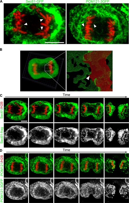
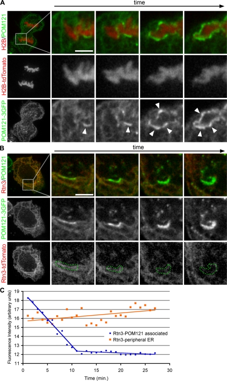
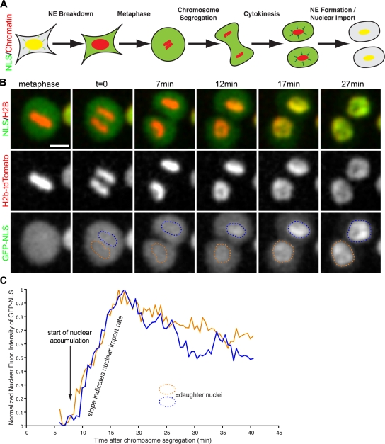
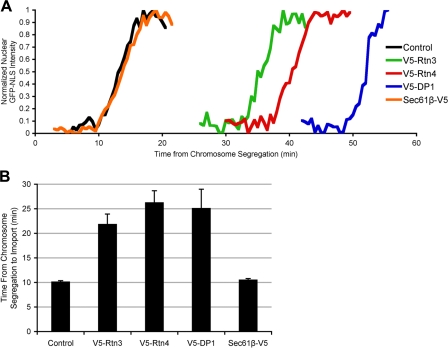
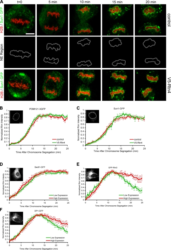
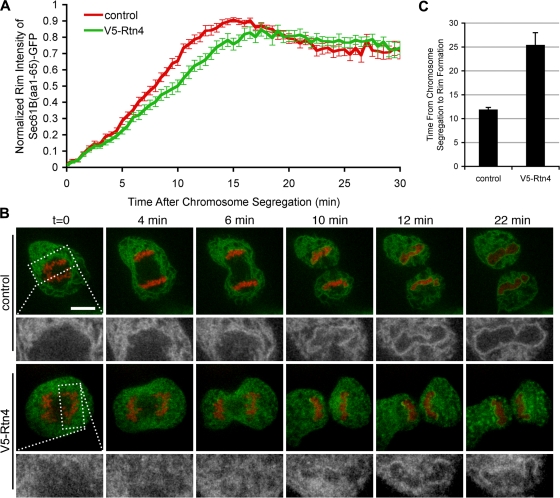
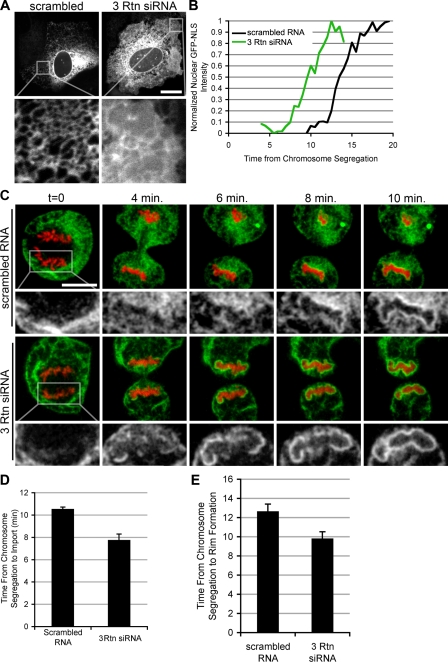
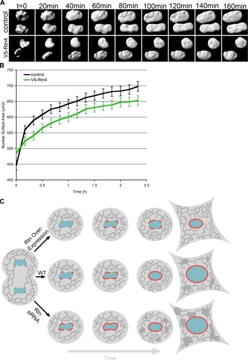
During mitosis in metazoans, segregated chromosomes become enclosed by the nuclear envelope (NE), a double membrane that is continuous with the endoplasmic reticulum (ER). Recent in vitro data suggest that NE formation occurs by chromatin-mediated reorganization of the tubular ER; however, the basic principles of such a membrane-reshaping process remain uncharacterized. Here, we present a quantitative analysis of nuclear membrane assembly in mammalian cells using time-lapse microscopy. From the initial recruitment of ER tubules to chromatin, the formation of a membrane-enclosed, transport-competent nucleus occurs within approximately 12 min. Overexpression of the ER tubule-forming proteins reticulon 3, reticulon 4, and DP1 inhibits NE formation and nuclear expansion, whereas their knockdown accelerates nuclear assembly. This suggests that the transition from membrane tubules to sheets is rate-limiting for nuclear assembly. Our results provide evidence that ER-shaping proteins are directly involved in the reconstruction of the nuclear compartment and that morphological restructuring of the ER is the principal mechanism of NE formation in vivo.


Figures

Figure 1.
Figure 1.
ER tubules bind and collapse on chromatin in vivo. U2OS cells were transfected with GFP fusion proteins and H2B-tdTomato, then imaged through mitosis with real-time spinning-disk confocal microscopy. (A) Large images of the initial interactions of Sec61-GFP and POM121-3GFP are shown. (B) z stacks were acquired every 30 s and initial membrane–chromatin contact points were characterized by 3D reconstruction. Arrowheads indicate initial membrane–chromatin contact. (C) Sec61-GFP was imaged every 5 s to monitor ER dynamics. (D) POM121-3GFP was imaged every 10 s to monitor the redistribution from the ER to the forming NE. Bars, 10 μm.
Figure 2.
Figure 2.
Rtn3 is removed from NE-forming tubules. (A) U2OS cells were transfected with POM121-3GFP and H2B-tdTomato to monitor the accumulation of POM121 at the forming NE. Arrowheads indicate forming NE. (B) U2OS cells were transfected with POM121-3GFP and Rtn3-tdTomato to monitor the dynamic localization of these proteins at the forming NE. Bars, 5 μm. (C) Intensity threshold selections of accumulating POM121 were used to define the regions of NE formation (dotted green regions in B). Fluorescence intensity of Rtn3-tdTomato was measured in these regions through NE formation (blue circles); intensity was also measured in the peripheral ER (orange squares). Cells were imaged every 30 s.
Figure 3.
Figure 3.
Assay developed to measure NE-formation kinetics. (A) Schematic of the dynamic localization of the reporter genes for nuclear localization (NLS) and chromatin during mitosis. Yellow represents the colocalization of NLS (green) and chromatin (red) in the intact nucleus. (B) U2OS were transfected with NLS fused to 3GFPs (GFP-NLS) and H2B-tdTomato, then imaged every 30 s through mitosis with live-cell confocal microscopy. Bar, 20 μm. (C) After chromosome segregation, regions over chromatin were selected, and GFL-NLS intensity was measured over time. Time = 0 corresponds to when the chromatid clusters have completely separated, and blue and orange traces correspond to the nuclear influx of GFP-NLS in each daughter nucleus.
Figure 4.
Figure 4.
ER tubule-shaping proteins delay NE formation. U2OS cells were transfected with GFP-NLS and H2B-tdTomato (control), with V5-tagged ER-shaping protein (Rtn3, Rtn4, or DP1), or with the V5-tagged ER protein Sec61β, then NE formation was analyzed. (A) Representative traces of nuclear GFP-NLS intensity during NE formation are shown. (B) Mean times from chromosome segregation to the onset of nuclear accumulation are plotted with standard error bars.
Figure 5.
Figure 5.
NE protein recruitment is not delayed by Rtn4 overexpression. A real-time microscopic assay was established to measure the recruitment of GFP fusion proteins to the forming NE. (A) U2OS cells were transfected with H2B-tdTomato and Sun1-GFP with or without the overexpression of V5-Rtn4 and imaged every 30 s through mitosis. A thin region surrounding the chromatin (NE region) was selected to measure the intensity of Sun1-GFP at the forming NE. (B) Fluorescence intensity of POM121-3GFP was measured at the NE region in control cells (red) or cells expressing V5-Rtn4 (green) starting at the onset of chromosome segregation, then normalized, averaged, and plotted with standard error bars. (C) Cells expressing Sun1-GFP were analyzed as in B. (D) U2OS cells were grouped for high (red) and low (green) expression of GFP-Sec61, and fluorescence intensity was measured at the forming NE as in A. (E) Cells expressing GFP-Rtn3 were analyzed as in D. (F) Cells expressing DP1-GFP were analyzed as in D. Interphase localization of each GFP fusion protein is shown in the insets in B–F. Bars: (A) 10 μm; (B) 5 μm.
Figure 6.
Figure 6.
NE rim formation is delayed by the overexpression of Rtn4. U2OS cells were transfected with Sec61-GFP and H2B-tdTomato, and imaged every 30 s in real time with confocal microscopy to quantify the time required for membrane rim formation. (A) Recruitment assay described in Fig. 6 was used to measure the recruitment of Sec61-GFP in control cells (red) and cells overexpressing Rtn4 (green). (B) Control cells and cells expressing V5-Rtn4 were imaged to focus on the membrane dynamics at the forming NE (high-magnification view in the bottom rows). (C) Time from chromosome segregation to nuclear rim formation was measured and averaged. Error bars represent standard error.
Figure 7.
Figure 7.
Reticulon knockdown accelerates NE formation. (A) U2OS cells were transfected with Sec61-GFP and scrambled RNA oligos or siRNA oligos directed against Rtn1, Rtn3, and Rtn4 (3 Rtn siRNA). ER morphology was then analyzed with confocal microscopy. Bar, 20 μm. (B) Cells transfected with scrambled RNA or 3 Rtn siRNA were analyzed using the NE formation kinetics assay as in Fig. 5. (C) Nuclear rim was analyzed as in Fig. 6, with cells transfected with either scrambled RNA or 3 Rtn siRNA. (D) The time of NE formation was averaged for cells transfected with scrambled RNA and 3 Rtn siRNA. Error bars represent standard error. (E) Time from chromosome segregation to rim formation was averaged. Error bars represent standard error.
Figure 8.
Figure 8.
Nuclear expansion is inhibited by Rtn4. U2OS cells were transfected with GFP-NLS and imaged every 10 min in 3D from mitosis through G1 by acquiring z stacks using confocal microscopy. (A) Nuclear surfaces were reconstructed from z stacks for control cells and cells overexpressing V5-Rtn4. (B) The surface areas of expanding nuclei were synchronized starting after nuclear accumulation of GFP-NLS and then averaged. Error bars indicate standard error. (C) Schematic shows the affects of ER-shaping protein concentration on NE formation and expansion, where the ER is represented in dark gray, chromatin is represented in blue, and red represents the forming NE.

References

    1. Anderson, D.J., and M.W. Hetzer. 2007. Nuclear envelope formation by chromatin-mediated reorganization of the endoplasmic reticulum. Nat. Cell Biol. 9:1160–1166. - PubMed
    1. Anderson, D.J., and M.W. Hetzer. 2008. The life cycle of the metazoan nuclear envelope. Curr. Opin. Cell Biol. 20:386–392. - PMC - PubMed
    1. Audhya, A., A. Desai, and K. Oegema. 2007. A role for Rab5 in structuring the endoplasmic reticulum. J. Cell Biol. 178:43–56. - PMC - PubMed
    1. Boman, A.L., M.R. Delannoy, and K.L. Wilson. 1992. GTP hydrolysis is required for vesicle fusion during nuclear envelope assembly in vitro. J. Cell Biol. 116:281–294. - PMC - PubMed
    1. Collas, P., and D. Poccia. 2000. Membrane fusion events during nuclear envelope assembly. Subcell. Biochem. 34:273–302. - PubMed

Publication types

MeSH terms