. 2008 Sep 8;182(5):937-50.
doi: 10.1083/jcb.200801152.

Cardiolipin defines the interactome of the major ADP/ATP carrier protein of the mitochondrial inner membrane

Affiliations

Cardiolipin defines the interactome of the major ADP/ATP carrier protein of the mitochondrial inner membrane

Steven M Claypool et al. J Cell Biol. .

Erratum in

  • J Cell Biol. 2012 Jun 25;197(7):1029

Abstract

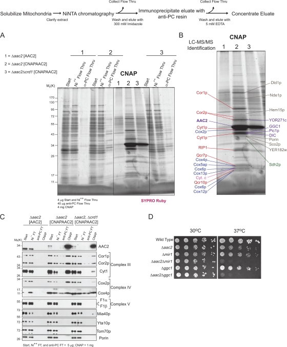
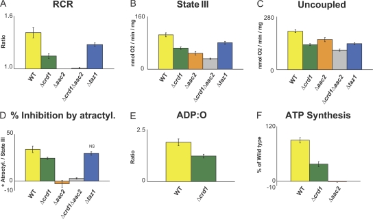
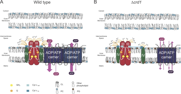
Defined mutations in the mitochondrial ADP/ATP carrier (AAC) are associated with certain types of progressive external ophthalmoplegia. AAC is required for oxidative phosphorylation (OXPHOS), and dysregulation of AAC has been implicated in apoptosis. Little is known about the AAC interactome, aside from a known requirement for the phospholipid cardiolipin (CL) and that it is thought to function as a homodimer. Using a newly developed dual affinity tag, we demonstrate that yeast AAC2 physically participates in several protein complexes of distinct size and composition. The respiratory supercomplex and several smaller AAC2-containing complexes, including other members of the mitochondrial carrier family, are identified here. In the absence of CL, most of the defined interactions are destabilized or undetectable. The absence of CL and/or AAC2 results in distinct yet additive alterations in respiratory supercomplex structure and respiratory function. Thus, a single lipid can significantly alter the functional interactome of an individual protein.


Figures

Figure 1.
Figure 1.
Disorganization of AAC complexes in the absence of CL. (A) 100 μg of 1.5% (wt/vol) digitonin extracts from mitochondria derived from the indicated strains were resolved by 2D BN/SDS-PAGE and AAC-complexes revealed by immunoblot. n = 3. (B) 25 μg of each subcellular fraction was immunoblotted for the indicated subcellular organelle. n = 2. (C) Steady-state expression was determined from whole cell extracts (5 and 10 μl) by immunoblotting for AAC (bottom), with Taz1p (middle) and Tom70p (top) serving as loading controls. n = 3. (D) CNAPAAC2 assembles in similar complexes as untagged AAC2. n = 3 (E) Serial dilutions of the strains indicated at the left were spotted onto YP medium with dextrose or ethanol–glycerol as the carbon source and incubated at 30°C for 3 d. n = 3. Asterisk highlights cross-reaction with porin of the AAC antiserum.
Figure 2.
Figure 2.
1D analysis of the AAC2 interactome with and without CL. (A) After CNAP of extracts derived from the indicated mitochondria, the final concentrated eluates were resolved by 10–15% SDS-PAGE. (B) SYPRO Ruby detected bands were extracted, digested with trypsin, and proteins identified by LC–MS/MS. Respiratory complex III and IV subunits are labeled in red and blue, respectively, and members of the mitochondrial carrier family in purple. (C) After CNAP, the final concentrated eluates were resolved by SDS-PAGE and immunoblotted for the indicated mitochondrial proteins. n = 3. (D) Serial dilutions of the strains indicated at the left were spotted onto YP medium with dextrose as the carbon source and incubated at the indicated temperature for 3 d.
Figure 3.
Figure 3.
2D BN/SDS-PAGE analysis of the AAC2 interactome with and without CL. (A–C) After CNAP, the final concentrated eluates were resolved in the first dimension by 6–16% BN-PAGE and in the second dimension by 12–18% SDS-PAGE. SYPRO Ruby detected bands were extracted, digested with trypsin, and proteins identified by LC–MS/MS. Respiratory complex III and IV subunits are labeled in red and blue, respectively, and members of the mitochondrial carrier family in purple. The pink circle highlights contaminating catalase (verified by LC–MS/MS) from the BN-PAGE molecular weight markers. (D–F) After CNAP, the final concentrated eluates were resolved by 2D BN/SDS-PAGE and immunoblotted for the indicated mitochondrial proteins. n = 3. The sources of the purified extracts were (A and D) Δaac2[AAC2], (B and E) Δaac2[CNAPAAC2], and (C and F) Δaac2Δcrd1[CNAPAAC2].
Figure 4.
Figure 4.
The largest AAC2-containing complex requires the presence of respiratory supercomplexes and is stabilized by CL. (A) Endogenous AAC2 coIPs with the respiratory supercomplex. An anti-complex IV IP was performed on digitonin extracts from the indicated mitochondria. 1% of the starting material and final flow-thru versus 100% of the preclear and IP beads post-washing were immunoblotted for the indicated mitochondrial proteins. n = 3. (B) Endogenous respiratory subunits coIP with AAC2. As in (A), except that 0.5% of the starting material and final flow-thru were analyzed. n = 3. (C) AAC2 and respiratory supercomplexes interact in organello. The anti-AAC2 IP was as in (B), except the preclear was omitted and the amount of protein analyzed is expressed as per mitochondrial source. n = 3. (D) wt or rho– mitochondrial extracts were resolved by 2D BN/SDS-PAGE and immunoblotted for the indicated mitochondrial proteins. n = 3. (E) After IP of wt digitonin extracts with normal rabbit serum or the anti-complex IV antiserum, the concentrated nonbinding flow-thrus were resolved by 2D BN/SDS-PAGE and immunoblotted for the indicated mitochondrial proteins. n = 3.
Figure 5.
Figure 5.
AAC2 and CL are required for the normal configuration of the respiratory supercomplexes. (A) Steady-state expression of assorted proteins (5, 10, and 20 μg protein) in the indicated mitochondria as assessed by immunoblot. n = 3. (B) After steady-state labeling with 32Pi, phospholipids were extracted from the indicated strains, separated by TLC, and revealed by phosphorimaging. The relative abundance of each phospholipid is expressed as a percentage of the total phospholipid in each strain (mean ± SEM, n = 3). Asterisks indicate significant accumulation of the designated phospholipid for a given mutant strain relative to wt (P ≤ 0.013) as determined by one-way anova, with Holm–Sidak pairwise comparisons. (C) Mitochondrial extracts from the indicated strains were resolved by both 1D BN-PAGE and 2D BN/SDS-PAGE, performed in parallel, and immunoblots performed for complex III (Cyt1 and Cor2p), complex IV (Cox2p), and complex V (F1β). The migrations of the III2IV2, III2IV, III2, IV, Vdimer, and Vmonomer supercomplexes are indicated, where appropriate, in the 1D BN-PAGE immunoblots. n = 4.
Figure 6.
Figure 6.
AAC2 and CL are required for the optimal functioning of the respiratory supercomplexes. 200 μg mitochondria were incubated in respiration buffer at 25°C and oxygen consumption was monitored in the presence of 5 mM succinate. (A) RCRs, (B) state III respiratory rates in the presence of 50 μM ADP, (C) uncoupled respiratory rates in the presence of 10 μM CCCP, (D) percent inhibition by 40 μM atractyloside compared with state III rates (mean ± SEM). (E) ADP:O ratios in the presence of 2 mM NADH, (F) ATP production (percentage of wt) by complex V in the presence of 5 mM NADH, 2 mM ADP, and 10 μM adenylate kinase inhibitor, Ap5A (mean ± SEM). Aliquots taken at 30, 60, 90, 120, and 150 s after addition of 2 mM ADP were extracted with PCA and ATP levels measured. Unless indicated by “NS”, all differences compared with wt are statistically significant (P ≤ 0.05) by t test. n = 6–11 (A–D), n = 5–9 (E), and n = 4 (F).
Figure 7.
Figure 7.
Model for how CL increases the efficiency of oxidative phosphorylation. (A) CL, the “green” phospholipid, not only facilitates cyt c (blue squares) transport between complexes III (red ovals) and IV (dark green ovals) by stabilizing the III2IV2+AAC2 supercomplex, but additionally stimulates AAC (six membrane domains represented by blue cylinders) activity by placing it in an electrochemical bath provided by the proton-coupled electron transport activity of complexes III and IV. (B) In the absence of CL, the net distance traveled by cyt c between complexes III and IV is increased and the potential benefits on AAC2 function by associating with the respiratory supercomplexes is lost, resulting in a reduced efficiency of OXPHOS. Not drawn to scale.

References

    1. Beyer, K., and B. Nuscher. 1996. Specific cardiolipin binding interferes with labeling of sulfhydryl residues in the adenosine diphosphate/adenosine triphosphate carrier protein from beef heart mitochondria. Biochemistry. 35:15784–15790. - PubMed
    1. Boumans, H., L.A. Grivell, and J.A. Berden. 1998. The respiratory chain in yeast behaves as a single functional unit. J. Biol. Chem. 273:4872–4877. - PubMed
    1. Brandner, K., D.U. Mick, A.E. Frazier, R.D. Taylor, C. Meisinger, and P. Rehling. 2005. Taz1, an outer mitochondrial membrane protein, affects stability and assembly of inner membrane protein complexes: implications for Barth Syndrome. Mol. Biol. Cell. 16:5202–5214. - PMC - PubMed
    1. Chicco, A.J., and G.C. Sparagna. 2007. Role of cardiolipin alterations in mitochondrial dysfunction and disease. Am. J. Physiol. Cell Physiol. 292:C33–C44. - PubMed
    1. Claypool, S.M., J.M. McCaffery, and C.M. Koehler. 2006. Mitochondrial mislocalization and altered assembly of a cluster of Barth syndrome mutant tafazzins. J. Cell Biol. 174:379–390. - PMC - PubMed

Publication types

MeSH terms

Substances