. 2008 Sep 23;105(38):14615-9.
doi: 10.1073/pnas.0803652105. Epub 2008 Sep 8.

Gender differences in human cortical synaptic density

Affiliations

Gender differences in human cortical synaptic density

L Alonso-Nanclares et al. Proc Natl Acad Sci U S A. .

Abstract

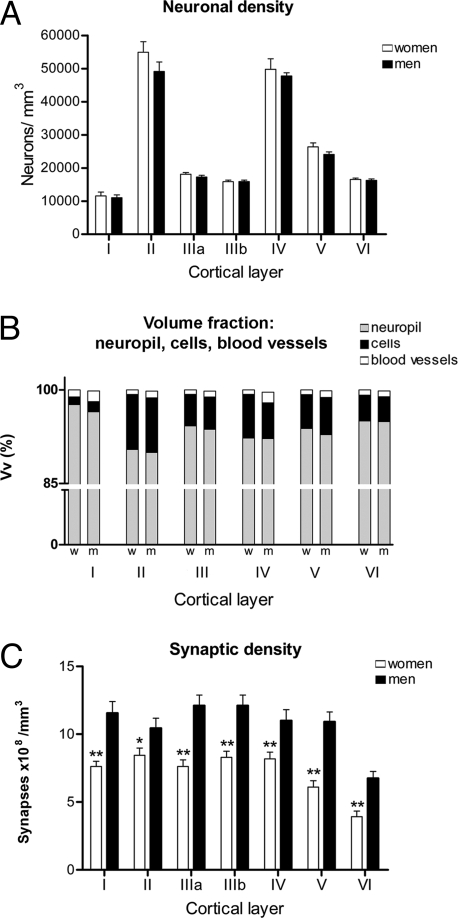
Certain cognitive functions differ in men and women, although the anatomical and functional substrates underlying these differences remain unknown. Because neocortical activity is directly related with higher brain function, numerous studies have focused on the cerebral cortex when searching for possible structural correlates of cognitive gender differences. However, there are no studies on possible gender differences at the synaptic level. In the present work we have used stereological and correlative light and electron microscopy to show that men have a significantly higher synaptic density than women in all cortical layers of the temporal neocortex. These differences may represent a microanatomical substrate contributing to the functional gender differences in brain activity.


Conflict of interest statement

The authors declare no conflict of interest.

Figures

Fig. 1.
Fig. 1.
Graphs of the neuronal densities, volume fraction (Vv), synaptic densities, and synapses per neurons in men and women in each cortical layer. (A) Graph showing the neuronal densities (mean ± SEM) in each cortical layer demonstrating that there are no significant differences between men and women. (B) Comparison of the Vv between men and women, calculated for the neuropil, cell bodies (including those from glia and neurons), and blood vessels in each cortical layer. Note that the neuropil represents between 90% and 98% of the volume, for which no significant differences were found between men and women. (C) Graph showing a comparison of synaptic density (mean ± SEM) between men and women in each cortical layer. w, women; m, men. *, P < 0.05; **, P < 0.01.
Fig. 2.
Fig. 2.
Electron micrographs to illustrate the ultrastructure of the human temporal neocortex. (A and B) Low-power electron micrographs showing the neuropil from layer IIIb of the temporal neocortex from a woman (A) and a man (B). Some synapses are indicated by arrows, and the asterisks illustrate two dendritic spines that are also shown at higher magnification in Fig. S3. (C and D) High-power electron micrographs showing the two major morphological types of synapses in the neuropil. Asymmetric synapses (arrows) had a prominent postsynaptic density, whereas symmetric synapses (arrowhead) had a thin postsynaptic density. de, dendritic shaft; ds, dendritic spines; T, axon terminals. [Scale bar (in D): 0.9 μm for A and B and 0.4 μm for C and D.]

Similar articles

Cited by

References

    1. Linn MC, Petersen AC. Emergence and characterization of sex differences in spatial ability: A meta-analysis. Child Dev. 1985;56:1479–1498. - PubMed
    1. Kimura D. Sex and Cognition. Cambridge, MA: MIT Press; 2000.
    1. Bocklandt S, Vilain E. Sex differences in brain and behavior: Hormones versus genes. Adv Genet. 2007;59:245–266. - PubMed
    1. Cosgrove KP, Mazure CM, Staley JK. Evolving knowledge of sex differences in brain structure, function, and chemistry. Biol Psychiatry. 2007;62:847–855. - PMC - PubMed
    1. Davies W, Wilkinson LS. It is not all hormones: Alternative explanations for sexual differentiation of the brain. Brain Res. 2006;1126:36–45. - PubMed

Publication types