. 2008 Sep 16;105(37):14028-33.
doi: 10.1073/pnas.0804358105. Epub 2008 Sep 8.

IL-21 signaling is critical for the development of type I diabetes in the NOD mouse

Affiliations

IL-21 signaling is critical for the development of type I diabetes in the NOD mouse

Rosanne Spolski et al. Proc Natl Acad Sci U S A. .

Abstract

IL-21 is a pleiotropic type I cytokine that shares the common cytokine receptor gamma chain and plays important roles for normal Ig production, terminal B cell differentiation to plasma cells, and Th17 differentiation. IL-21 is elevated in several autoimmune diseases, and blocking its action has attenuated disease in MRL/lpr mice and in collagen-induced arthritis. The diabetes-associated Idd3 locus is at the Il2/Il21 locus, and elevated IL-21 was observed in the nonobese diabetic (NOD) mouse and suggested to contribute to diabetes by augmenting T cell homeostatic proliferation. To determine the role of IL-21 in diabetes, Il21r-knockout (KO) mice were backcrossed to NOD mice. These mice were devoid of lymphocytic infiltration into the pancreas, and only 1 of 20 animals had an elevated glucose compared with 60% of NOD mice on a wild-type (WT) background. Although TCR and Treg-related responses were normal, these mice had reduced Th17 cells and significantly higher levels of mRNAs encoding members of the Reg (regenerating) gene family whose transgenic expression protects against diabetes. Our studies establish a critical role for IL-21 in the development of type I diabetes in the NOD mouse, with obvious potential implications for type I diabetes in humans.


Conflict of interest statement

Conflict of interest statement: R.S. and W.J.L. are inventors on patents and patent applications related to IL-21.

Figures
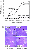
Fig. 1.
Fig. 1.
NOD/Il21r-KO mice do not develop diabetes or insulitis. (A) Il21r-KO mice were bred onto the NOD background for seven generations. Blood glucose levels were measured weekly in 51 NOD/WT and 20 NOD/Il21r-KO littermates starting at the age of 10 weeks, and glucose levels >250 mg/dl were scored as diabetic. Twenty-two NOD/Il21r+/− mice were also analyzed up to 20 weeks of age, at which point they were killed. In addition, blood glucose levels were measured in all subsequently killed KO mice used in this work and were found to be in the normal range (<200 mg/dl). (B) Representative pancreatic sections from 12-week-old NOD/WT and NOD/Il21r-KO mice showing typical islet infiltration only in the WT pancreas (Left). (Magnification: Upper, ×100; Lower, ×200.) Four additional NOD/WT and NOD/Il21r-KO mice were examined, with results similar to those shown in the figure. H&E stain was used. (Scale bars: 100 μm.)
Fig. 2.
Fig. 2.
Normal lymphoid cellularity and activation markers in NOD mice. (A and B) Splenic T cell populations and pancreatic lymph node cellularity were quantified by flow cytometry (n = 12 for NOD/WT, n = 12 for NOD/Il21r-KO, n = 6 for BALB/c WT, n = 6 for BALB/cIl21r-KO); mean ± SEM. (A) Tabulation of data. (B) Representative flow cytometric profiles. (C) Splenic T cells were analyzed by flow cytometry for the activation markers CD62L, CD44, and IL-2Rβ. Representative FACS profiles from one of three independent experiments are shown. (D) CD4+ T cells were isolated and stimulated either with or without anti-CD3/anti-CD28 for 6 h, at which time IL-21 and IFN-γ mRNA were analyzed by quantitative RT-PCR. The results were normalized relative to expression of Rpl7. Shown are mean ± SD from three mice in each group; results are representative of three independent experiments. (E) CD4+ T cells (2 × 10⁶ per ml) were stimulated with anti-CD3/anti-CD28 either in the presence or absence of IL-6 for 48 h, at which time supernatants were assayed by ELISA for either IL-21 or IFN-γ. Shown is the mean ± SD for results from three mice in each group.
Fig. 3.
Fig. 3.
T cell proliferative responses and Treg cell function are normal in NOD/Il21r-KO mice. (A and B) Splenocytes (A) or pancreatic lymph nodes (B) (106 per ml) were cultured in microwells with plate-bound anti-CD3 at the indicated concentrations for splenocytes and at 2 μg/ml for pancreatic lymph nodes for 48 h and were pulsed with [3H]thymidine for the last 8 h. No significant difference was observed in the proliferation of NOD/WT vs. NOD/Il21r-KO splenocytes or pancreatic lymph node cells. Shown is a representative experiment, with error bars indicating the SD for a group of three mice. (C) Splenic CD4+ or CD8+ T cells from WT or KO mice were stimulated with increasing concentrations of anti-CD3 in the presence or absence of irradiated T-depleted splenic APCs from either WT or KO mice for 48 h and were pulsed with [3H]thymidine for the last 8 h. Shown is a representative experiment, with error bars indicating the SD for a group of three mice. (D) Splenic and pancreatic LN Treg cells were quantitated by intracellular staining with FoxP3 Ab. Representative staining profiles are shown. Average percentages of FoxP3+ pancreatic lymph nodes of nine mice from three different experiments were as follows: NOD/WT, 3.6 ± 0.3; NOD/Il21r-KO, 3.9 ± 0.4. (E) CD4+ NOD/WT or NOD/Il21r-KO cells (5 × 10⁴ per well) were cultured with increasing numbers of purified splenic Tregs from either NOD/WT or NOD/Il21r-KO mice in the presence of irradiated syngeneic T-depleted APCs and anti-CD3 for 3 days with [3H]thymidine labeling during the last 8 h of culture. Shown is mean ± SD for results from three mice per group from a representative experiment.
Fig. 4.
Fig. 4.
IL-17 production is reduced in CD4+ T cells from pancreatic lymph nodes or spleen of NOD/Il21r-KO mice. (A) T cells from pancreatic lymph node or spleen were cultured with either TGF-β (2 ng/ml) + IL-6 (2 ng/ml) (Upper) or with IL-12 (10 ng/ml) (Lower) in plates with plate-bound anti-CD3 (2 μg/ml) + anti-CD28 (1 μg/ml) for 48 h. Cells were then stimulated with PMA + ionomycin in the presence of Golgi plug for 5 h and were intracellularly stained with IL-17A and IFN-γ mAbs. Shown is a representative profile of one of three mice from an experiment with three mice in each experimental group; three similar independent experiments were performed. (B) Culture supernatants were collected at 48 h, and levels of secreted IL-17 and IFN-γ were measured by ELISA. Shown is mean ± SD for results from three mice per group from a representative experiment. (C) CD4+ T cells from NOD/WT or NOD/Il21r-KO mice were stimulated under nonpolarizing conditions with anti-CD3 either in the absence or the presence of irradiated T-depleted splenic APC for 48 h, and culture supernatants were assayed for secreted IL-17 and IFN-γ by ELISA.
Fig. 5.
Fig. 5.
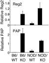
Il21r-KO pancreas expresses higher levels of two members of the Reg family of mRNAs. Pancreatic RNA from 6- to 8-week-old WT and Il21r-KO mice on either the C57BL/6 or the NOD background was prepared and subjected to analysis by quantitative real-time PCR. RT-PCR revealed significantly higher levels of Reg2 or PAP mRNA in Il21r-KO mice on either background. Shown is the mean ± SD for three mice in each experimental group. Three similar independent experiments were performed.

Similar articles

Cited by

References

    1. Powers AC. Diabetes Mellitus. New York: McGraw–Hill; 2005.
    1. Anderson M, Bluestone J. The NOD mouse: A model of immune dysregulation. Annu Rev Immunol. 2005;23:447–485. - PubMed
    1. Parrish-Novak J, et al. Interleukin 21 and its receptor are involved in NK cell expansion and regulation of lymphocyte function. Nature. 2000;408:57–63. - PubMed
    1. Spolski R, Leonard WJ. Interleukin-21: Basic biology and implications for cancer and autoimmunity. Annu Rev Immunol. 2008;26:57–79. - PubMed
    1. Noguchi M, et al. Interleukin-2 receptor γ chain mutation results in X-linked severe combined immunodeficiency in humans. Cell. 1993;73:147–157. - PubMed

Publication types

MeSH terms