. 2008 Sep 16;105(37):14175-80.
doi: 10.1073/pnas.0807163105. Epub 2008 Sep 8.

C-terminal peptides coassemble into Abeta42 oligomers and protect neurons against Abeta42-induced neurotoxicity

Affiliations

C-terminal peptides coassemble into Abeta42 oligomers and protect neurons against Abeta42-induced neurotoxicity

Erica A Fradinger et al. Proc Natl Acad Sci U S A. .

Abstract

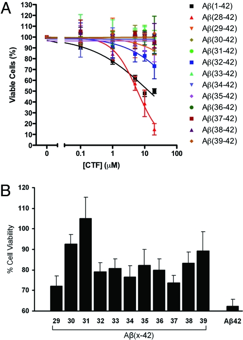
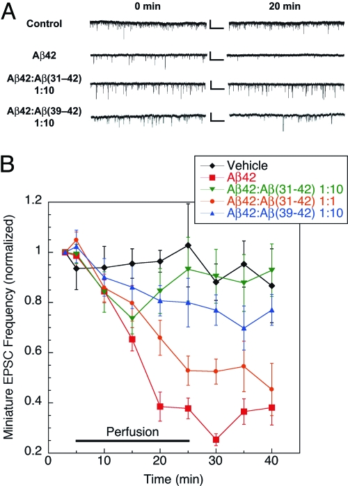
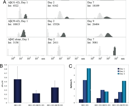
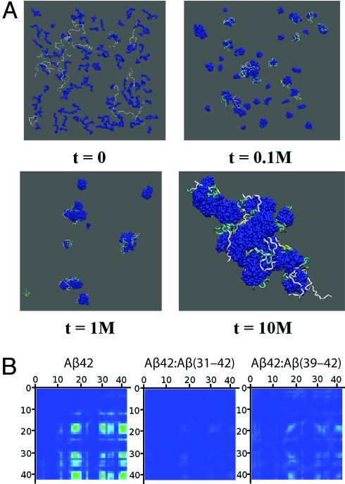
Alzheimer's disease (AD) is an age-related disorder that threatens to become an epidemic as the world population ages. Neurotoxic oligomers of Abeta42 are believed to be the main cause of AD; therefore, disruption of Abeta oligomerization is a promising approach for developing therapeutics for AD. Formation of Abeta42 oligomers is mediated by intermolecular interactions in which the C terminus plays a central role. We hypothesized that peptides derived from the C terminus of Abeta42 may get incorporated into oligomers of Abeta42, disrupt their structure, and thereby inhibit their toxicity. We tested this hypothesis using Abeta fragments with the general formula Abeta(x-42) (x = 28-39). A cell viability screen identified Abeta(31-42) as the most potent inhibitor. In addition, the shortest peptide, Abeta(39-42), also had high activity. Both Abeta(31-42) and Abeta(39-42) inhibited Abeta-induced cell death and rescued disruption of synaptic activity by Abeta42 oligomers at micromolar concentrations. Biophysical characterization indicated that the action of these peptides likely involved stabilization of Abeta42 in nontoxic oligomers. Computer simulations suggested a mechanism by which the fragments coassembled with Abeta42 to form heterooligomers. Thus, Abeta(31-42) and Abeta(39-42) are leads for obtaining mechanism-based drugs for treatment of AD using a systematic structure-activity approach.


Conflict of interest statement

The authors declare no conflict of interest.

Figures

Fig. 1.
Fig. 1.
Evaluation of CTF effect on neuronal cultures. CTFs at final nominal concentrations of 0.1–20 μM or mixtures of Aβ42:CTF at a 1:10 concentration ratio, respectively, were incubated with differentiated PC-12 cells. In A, Aβ42 (black squares) is shown for comparison. In B, the nominal concentration of Aβ42 is 5 μM. After 15 h of incubation, cell viability was measured by using the MTT assay. Cell culture medium containing DMSO in the same concentrations as used for peptide solubilization was used as a negative control, and 1 μM staurosporine was used as a positive control. The graphs show average data ± SD from at least three independent experiments, each performed with six wells per condition.
Fig. 2.
Fig. 2.
CTFs rescue mEPSCs in Aβ42-treated hippocampal neurons. Mouse primary hippocampal neurons were exposed to vehicle (n = 5), 3 μM Aβ42 (n = 8), 1:1 Aβ42:Aβ(31–42) (n = 9), 1:10 Aβ42:Aβ(31–42) (n = 10), or 1:10 Aβ42:Aβ(39–42) (n = 6) mixtures, and the frequency and amplitude of mEPSCs were measured. (A) Representative recording traces collected before (0 min) and 20 min after peptide perfusion. Calibration bars: 25 pA/1 sec. (B) Cells were perfused with vehicle for 5 min to establish baseline, and then with peptide solutions for an additional 20 min, and allowed to recover in vehicle solution for 15 min. The curves show the time dependence of mEPSC frequency after exposure to Aβ42 in the absence or presence of CTFs over 40 min.
Fig. 3.
Fig. 3.
CTF effect on Aβ42 assembly. (A) Representative distributions of Aβ42 in the absence or presence of CTFs immediately after preparation (Left), on the next day (Center), and after 7 or 9 days (Right). White bars represent P1 particles. Gray bars represent P2 or larger particles (in the case of Aβ42 alone). Days of measurement and the total scattering intensities in counts per second are shown in the upper left corner of each panel. Only intensities within the same row are directly comparable with each other. (B) Growth rates of P2 particles (dRH2/dt) in the absence or presence of CTFs. (C) Average number of intensity spikes per hour during the first 3 days of measurement in the absence or presence of CTFs.
Fig. 4.
Fig. 4.
Simulation of the interaction between Aβ42 and CTFs during oligomerization. (A) Configurations of 16 Aβ42 and 128 Aβ(31–42) molecules at different time frames measured at t simulation steps. CTFs are displayed in dark blue, and Aβ42 molecules are represented by their secondary structure: yellow ribbons, β-strands; blue tubes, turns; silver tubes, random coil. (B) Intermolecular contact maps of Aβ42 in the absence or presence of CTFs calculated for the highest Aβ42:CTF peptide number concentration ratio (1:8). The contact maps are oriented such that the contact strength between pairs of N-terminal residues is displayed at the top left corner and the contact strength between pairs of C-terminal residues is at the bottom right corner. The strength of the contact between two amino acids is color-coded from 0.0 (blue) to a maximal strength (red), corresponding to 30 contacts.

References

    1. Brookmeyer R, Johnson E, Ziegler-Graham K, Arrighi HM. Forecasting the global burden of Alzheimer's disease. Alzheimer's Dementia. 2007;3:186–191. - PubMed
    1. Alzheimer Association. 2008 Alzheimer's disease facts and figures. Alzheimer's Dementia. 2008;4:110–133. - PubMed
    1. Hardy JA, Higgins GA. Alzheimer's disease: The amyloid cascade hypothesis. Science. 1992;256:184–185. - PubMed
    1. Kirkitadze MD, Bitan G, Teplow DB. Paradigm shifts in Alzheimer's disease and other neurodegenerative disorders: The emerging role of oligomeric assemblies. J Neurosci Res. 2002;69:567–577. - PubMed
    1. Walsh DM, Selkoe DJ. Aβ oligomers—A decade of discovery. J Neurochem. 2007;101:1172–1184. - PubMed

Publication types

MeSH terms