. 2008 Sep 30;105(39):15172-7.
doi: 10.1073/pnas.0806717105. Epub 2008 Sep 8.

Physiological significance of a peripheral tissue circadian clock

Affiliations

Physiological significance of a peripheral tissue circadian clock

Katja A Lamia et al. Proc Natl Acad Sci U S A. .

Abstract

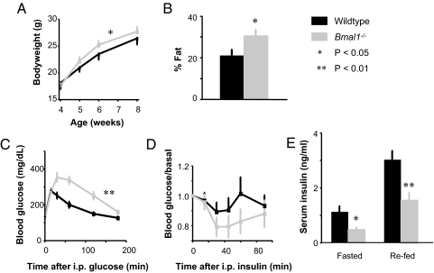
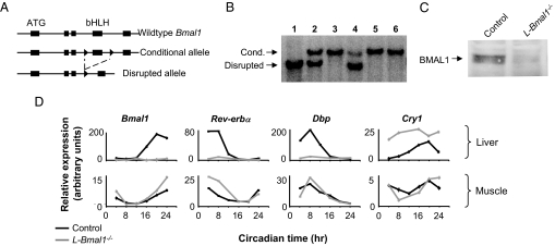
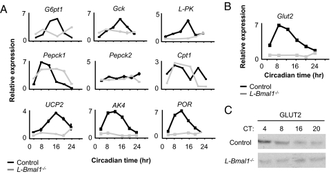
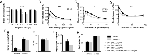
Mammals have circadian clocks in peripheral tissues, but there is no direct evidence of their physiological importance. Unlike the suprachiasmatic nucleus clock that is set by light and drives rest-activity and fasting-feeding cycles, peripheral clocks are set by daily feeding, suggesting that at least some contribute metabolic regulation. The liver plays a well known role in glucose homeostasis, and we report here that mice with a liver-specific deletion of Bmal1, an essential clock component, exhibited hypoglycemia restricted to the fasting phase of the daily feeding cycle, exaggerated glucose clearance, and loss of rhythmic expression of hepatic glucose regulatory genes. We conclude that the liver clock is important for buffering circulating glucose in a time-of-day-dependent manner. Our findings suggest that the liver clock contributes to homeostasis by driving a daily rhythm of hepatic glucose export that counterbalances the daily cycle of glucose ingestion resulting from the fasting-feeding cycle.


Conflict of interest statement

The authors declare no conflict of interest.

Figures

Fig. 1.
Fig. 1.
Glucose intolerance and abnormal energy balance in mice lacking Bmal1 function in all tissues. Shown are comparisons of Bmal1−/− mice and wild-type littermates (C57BL/6 × 129). (A) Bodyweight (ANOVA). (B) Total body fat content (t test). (C) Glucose tolerance (ANOVA); Zeitgeber time (ZT, in h) 4.5. (D) Insulin tolerance (ANOVA; the trend toward hypersensitivity was not significant); ZT 8.5 (in a 12-h light/12-h dark cycle). (E) Serum insulin concentration after overnight fast with or without refeeding (t test); ZT 2.5 and 4.5, respectively. Shown are mean and SEM of 7–9 mice of each genotype. *, P < 0.05; **, P < 0.01.
Fig. 2.
Fig. 2.
Liver-specific loss of circadian clock function. (A) Conditional Bmal1 allele and disruption by Cre recombinase. Boxes, exons; ATG, translation start site; bHLH, basic helix–loop—helix domain; triangles, loxP sites. (B) Liver-specific disruption of Bmal1 conditional allele: genomic Southern blot showing fragments diagnostic of the conditional or disrupted Bmal1 alleles, as marked. Lanes 1–4, liver genomic DNA. Lane 1, homozygous Bmal1 conditional, ubiquitous Cre; lane 2, heterozygous for disrupted allele; lane 3, homozygous Bmal1 conditional, no Cre; and lane 4, homozygous Bmal1 conditional, albumin-Cre. Lanes 5 and 6, genomic DNA from skeletal muscle and kidney, respectively, from same mouse as lane 4. (C) Loss of BMAL1 protein in the liver: anti-BMAL1 Western blot of protein extracts from livers of L-Bmal1−/− and littermate control mice, as indicated. (D) Liver-specific loss of molecular circadian rhythms: quantitative reverse-transcriptase PCR (Q-PCR) showing temporal expression profiles of Bmal1 and other clock-associated genes, as indicated, in liver and muscle of L-Bmal1−/− mice and littermate controls. Shown are the mean and SEM of triplicate assays (most error bars are too small to be seen at this scale). Dbp, D-site albumin promoter binding protein; Cry1, Cryptochrome 1.
Fig. 3.
Fig. 3.
Loss of rhythmic expression of clock-regulated metabolic genes in the livers of L-Bmal1−/− mice. (A) One-day temporal expression profiles of selected hepatic metabolic genes. Shown are the mean and SEM of Q-PCR triplicate assays (most error bars are too small to be seen at this scale). G6pt1, glucose-6-phosphate translocase 1; Gck, glucokinase; L-PK, liver pyruvate kinase; Pepck1, phosphoenolpyruvate carboxykinase 1; Pepck2, phosphoenolpyruvate carboxykinase 2; Cpt1, carnitine palmitoyltransferase 1; Ucp2, uncoupling protein 2; AK4, adenylate kinase 4; POR, P450 oxidoreductase. (B) One-day temporal expression profile of hepatic Glut2 (glucose transporter 2) transcript in the indicated genotypes. Shown are mean and SEM of Q-PCR triplicate assays (most error bars are too small to be seen at this scale). (C) One-day expression profile of GLUT2 protein. Shown is a Western blot of liver protein extracts obtained from mice of the indicated genotypes. CT, circadian time.
Fig. 4.
Fig. 4.
Hypoglycemia restricted to the fasting phase and exaggerated glucose clearance in mice with a liver-specific loss of circadian clock function. (A) Resting blood glucose of mice with the indicated genotypes measured across a 24-h cycle. The left half of the profile corresponds to the fasting phase of the daily behavioral cycle. The difference between genotypes over time is accounted for by the differences at fasting phase time-points ZT 4.5 and ZT 8.5. (B) Glucose tolerance; ZT 4.5. (C) Glucose tolerance after overnight fasting; ZT 4.5. (D) Insulin tolerance. The curves converge, with the significant difference between genotypes arising from the lower initial blood glucose in L-Bmal1−/− mice; ZT 8.5. (E) Serum insulin concentrations after mice were fasted overnight or fasted overnight and refed, as labeled. (F) Total body fat content. (G) Liver glycogen content. (H) Bodyweight at the indicated ages. For E–H, there were no significant differences between genotypes. Shown are the mean and SEM for 7–9 mice of each genotype. *, P < 0.05, ANOVA; **, P < 0.02, ANOVA; ***, P < 0.001, ANOVA; ∧, P < 0.05, Scheffé's post hoc analysis.

Comment in

References

    1. Young MW, Kay SA. Time zones: A comparative genetics of circadian clocks. Nat Rev Genet. 2001;2:702–715. - PubMed
    1. Ko CH, Takahashi JS. Molecular components of the mammalian circadian clock. Hum Mol Genet. 2006;15:R271–R277. - PubMed
    1. Abe M, et al. Circadian rhythms in isolated brain regions. J Neurosci. 2002;22:350–356. - PMC - PubMed
    1. Tosini G, Menaker M. Circadian rhythms in cultured mammalian retina. Science. 1996;272:419–421. - PubMed
    1. Balsalobre A, Damiola F, Schibler U. A serum shock induces circadian gene expression in cultured Rat-1 fibroblasts. Cell. 1998;93:929–937. - PubMed

Publication types