. 2008 Sep 16;105(37):14082-7.
doi: 10.1073/pnas.0804597105. Epub 2008 Sep 8.

Dicer-dependent endothelial microRNAs are necessary for postnatal angiogenesis

Affiliations

Dicer-dependent endothelial microRNAs are necessary for postnatal angiogenesis

Yajaira Suárez et al. Proc Natl Acad Sci U S A. .

Abstract

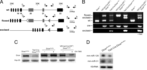
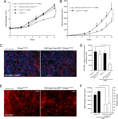
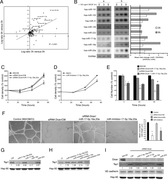
Posttranscriptional gene regulation by microRNAs (miRNAs) is important for many aspects of development, homeostasis, and disease. Here, we show that reduction of endothelial miRNAs by cell-specific inactivation of Dicer, the terminal endonuclease responsible for the generation of miRNAs, reduces postnatal angiogenic response to a variety of stimuli, including exogenous VEGF, tumors, limb ischemia, and wound healing. Furthermore, VEGF regulated the expression of several miRNAs, including the up-regulation of components of the c-Myc oncogenic cluster miR-17-92. Transfection of endothelial cells with components of the miR-17-92 cluster, induced by VEGF treatment, rescued the induced expression of thrombospondin-1 and the defect in endothelial cell proliferation and morphogenesis initiated by the loss of Dicer. Thus, endothelial miRNAs regulate postnatal angiogenesis and VEGF induces the expression of miRNAs implicated in the regulation of an integrated angiogenic response.


Conflict of interest statement

The authors declare no conflict of interest.

Figures

Fig. 1.
Fig. 1.
Conditional inactivation of Dicer in ECs. (A) Gene targeting strategy for endothelial-specific deletion of Dicer. PCR primers sets and fragment size expected are shown. (B) PCR genotyping analysis. (Top) Detection of floxed, excised, and WT fragments with primers a, b, and c. (Middle) Detection of Cre. (Bottom) Detection of excised fragment with primers b and c. (C) (Left) Dicer protein levels from ECs, isolated from 3-week-old Dicerflox/flox and Tie2-Cre;Dicerflox/flox mice. (Right) Dicer protein levels from EC isolated from 3-week-old Dicerflox/flox and VECad-Cre-ERT2;Dicerflox/flox mice. ECs were treated with OH-TMX or vehicle. (D) Reduction in miR-126 and -31 in ECs isolated from Tie2-Cre;Dicerflox/flox mice.
Fig. 2.
Fig. 2.
Endothelial Dicer inactivation reduces VEGF-induced angiogenesis. Ad-VEGF or Ad-eGFP was injected intradermally into the left and right ears, respectively, of 8-week-old mice. Frozen sections were stained for PECAM-1 (red) and counterstained with DAPI (blue). (A) Representative PECAM-1 staining showing reduced VEGF-induced angiogenesis in Tie2-Cre;Dicerflox/flox mice compared with Dicerflox/flox control mice. (B) Quantification of PECAM-1-positive structures (n = 6 per group). In B, for each animal, two to four images from five sections were quantified. Values are means ± SEM. *, P < 0.05, compared with Dicerflox/flox by Student's t test. (Scale bars: 100 μm.)
Fig. 3.
Fig. 3.
Endothelial Dicer inactivation reduces tumor angiogenesis and progression. (A and B) LLC cells were implanted intramuscularly into the calves of 8-week-old Dicerflox/flox or VECad-Cre-ERT2;Dicerflox/flox mice (A) or implanted s.c. in the dorsal flank of Dicerflox/flox and TMX-treated VECad-Cre-ERT2;DicerΔ/Δ mice (B), and tumor growth was assessed. n = 4 per group. P < 0.05 using a two-way ANOVA, followed by post hoc Bonferroni test. (C) Representative PECAM-1 staining (red) of cross-sections from tumors showing reduced tumor angiogenesis in VECad-Cre-ERT2;Dicerflox/flox mice. Tissues were counterstained with DAPI (blue). (D) Quantification of PECAM-1-positive structures in the tumors of the indicated groups of mice. For each tumor, two to four images from three sections from two different tumor pieces were quantified. (E) Representative whole-mount PECAM-1 staining from tumors isolated from dorsal flank showing reduced tumor-induced angiogenesis in VECad-Cre-ERT2;Dicerflox/flox mice. (F) Quantification of microvessel area and branch points, left and right bars for each group, respectively; data are from three to five images per sample. *, P < 0.05, compared with Dicerflox/flox by Student's t test. All data are mean ± SEM. (Scale bars: C and E, 100 μm.)
Fig. 4.
Fig. 4.
VEGF regulation of miRNA expression in ECs. (A) Data are the Log2 ratio of miRNA expression 3 and 9 h after VEGF treatment. hsa-miR-191, hsa-miR-155, hsa-miR-31, hsa-miR-126, hsa-miR-222, hsa-miR-7-5p, hsa-miR-18a, and hsa-miR-20a are highlighted in grey. (B) Confirmation and quantification of miRNA expression by Northern blotting. 5S rRNA served as a loading control. a and b denote samples from two different isolations of HUVECs. (C) Dicer knockdown decreases EC growth, an effect rescued by hsa-miR-17-5p, hsa-miR-18a, and hsa-miR-20a. EC (EA.hy.926 cells) were transfected with control nonsilencing and control mimic sequences (NS/CM) or Dicer siRNA in the presence of CM (siRNA Dicer/CM) or Dicer siRNA in the presence of a mix of specific mimics. (D) Targeting components of the miR-17-92 cluster reduces EC growth. ECs were transfected as above with control inhibitor sequence (CI) or a mixture of specific miRNA inhibitors and cell density was quantified. Data are expressed as percentage of control at day 0. (E). ECs were transfected as described in C for 48 h. Cells were incubated in medium without serum for 12 h (time = 0 h) and subsequently incubated in the presence of VEGF for 24 or 48 h. siRNA Dicer + miR-17-5p;18a;20a indicates cells that were initially transfected with Dicer siRNA for 48 h then transfected with miRNA mimics at the time of VEGF treatment. At the indicated time points, cell number was quantified. Data are expressed as percentage of control (NS/CM). (F) HUVECs were transfected as indicated in C. After 60 h, cells were seeded on a growth factor-reduced Matrigel in the presence of FBS (0.5%) and VEGF (100 ng/ml). Cumulative sprout length of capillary-like structures was quantified after 16 h. Data are from n = 3 experiments. (Magnification: ×10.) (G and H) Western blot analysis of Tsp1 levels in ECs transfected with CM, a mix of specific mimics, or individual specific mimics as indicated in G or transfected with CI, a mix of specific miRNA inhibitors, or individual specific miRNA inhibitors as indicated in H. Hsp-90 was used as loading control. (I) Western blot analysis of Dicer and Tsp1 expression levels in ECs transfected as indicated in C and in the presence of Dicer siRNA. VE-cadherin served as an EC marker and Hsp-90 served as loading control. Data are reported as the mean ± SEM. * and #, P < 0.05, by Student's t test.

References

    1. He L, Hannon GJ. MicroRNAs: Small RNAs with a big role in gene regulation. Nat Rev Genet. 2004;5:522–531. - PubMed
    1. Vasudevan S, Tong Y, Steitz JA. Switching from repression to activation: microRNAs can up-regulate translation. Science. 2007;318:1931–1934. - PubMed
    1. Bernstein E, et al. Dicer is essential for mouse development. Nat Genet. 2003;35:215–217. - PubMed
    1. Yang WJ, et al. Dicer is required for embryonic angiogenesis during mouse development. J Biol Chem. 2005;280:9330–9335. - PubMed
    1. Harfe BD, McManus MT, Mansfield JH, Hornstein E, Tabin CJ. The RNaseIII enzyme Dicer is required for morphogenesis but not patterning of the vertebrate limb. Proc Natl Acad Sci USA. 2005;102:10898–10903. - PMC - PubMed

Publication types

MeSH terms

LinkOut - more resources