. 2008 Sep 16;105(37):14181-6.
doi: 10.1073/pnas.0803893105. Epub 2008 Sep 8.

Intraretinal signaling by ganglion cell photoreceptors to dopaminergic amacrine neurons

Affiliations

Intraretinal signaling by ganglion cell photoreceptors to dopaminergic amacrine neurons

Dao-Qi Zhang et al. Proc Natl Acad Sci U S A. .

Abstract

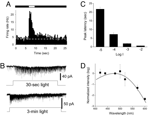
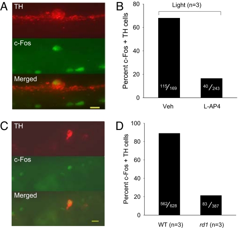
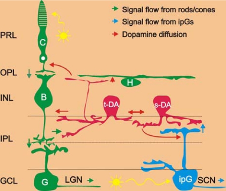
Retinal dopaminergic amacrine neurons (DA neurons) play a central role in reconfiguring retinal function according to prevailing illumination conditions, yet the mechanisms by which light regulates their activity are poorly understood. We investigated the means by which sustained light responses are evoked in DA neurons. Sustained light responses were driven by cationic currents and persisted in vitro and in vivo in the presence of L-AP4, a blocker of retinal ON-bipolar cells. Several characteristics of these L-AP4-resistant light responses suggested that they were driven by melanopsin-expressing intrinsically photosensitive retinal ganglion cells (ipRGCs), including long latencies, marked poststimulus persistence, and a peak spectral sensitivity of 478 nm. Furthermore, sustained DA neuron light responses, but not transient DA neuron responses, persisted in rod/cone degenerate retinas, in which ipRGCs account for virtually all remaining retinal phototransduction. Thus, ganglion-cell photoreceptors provide excitatory drive to DA neurons, most likely by way of the coramification of their dendrites and the processes of DA neurons in the inner plexiform layer. This unprecedented centrifugal outflow of ganglion-cell signals within the retina provides a novel basis for the restructuring of retinal circuits by light.


Conflict of interest statement

The authors declare no conflict of interest.

Figures

Fig. 1.
Fig. 1.
Transient and sustained light responses in dopaminergic amacrine neurons. (A and B) Extracellular loose patch recordings from a representative transient DA neuron (A) and representative sustained DA neuron (B). (Upper) Spike recordings made before and during L-AP4 application (50 μM). (Lower) Corresponding peri-stimulus time histograms (PSTH, bin width: 300 ms in A, 500 ms in B) before and during L-AP4 application. Bars indicate light on (open bar, duration 3 s, intensity −2 log I) and light off (filled bar). Dashed lines indicate prestimulus baseline. (C–E) Whole-cell voltage clamp recordings of light responses from transient and sustained DA neurons. Light-evoked excitatory inward currents in a transient DA neuron were blocked (C), whereas those in a sustained DA neuron were resistant to 75 μM L-AP4 (D), and L-AP4-resistant currents in a sustained DA neuron were blocked by 40 μM DNQX (E). Stimulus duration 3 s, intensity −1 log I in C and −2 log I in D and E.
Fig. 2.
Fig. 2.
L-AP4 resistant DA neuron light responses have characteristics of melanopsin phototransduction. All experiments were performed in the presence of L-AP4. (A) Average PSTH of DA neuron discharge before, during, and after light stimulus recorded by loose patch (bin width: 300 ms, n = 6, intensity −2 log I). (B) Typical whole-cell recordings show light-induced inward current that persisted throughout a 30-s light pulse (upper traces, −4 log I) and a 3-min light pulse (lower traces, −4 log I). (C) The latency of the peak light-induced current was long in duration and decreased as light intensity increased. (D) Lambda-max of the L-AP4-resistant light responses as determined by the method of ref. . For additional details see SI Methods. Mean data from 4 DA neurons were fit with Lamb's nomogram.
Fig. 3.
Fig. 3.
L-AP4 resistant light responses of DA neurons persist in rod/cone degenerate retinas. (A) DA neuron loose patch recording from a 13-month-old rd1 mouse retina showing light response and persistence in the presence of 50 μM L-AP4. Representative examples of recordings made before (Left) and during (Right) L-AP4 application. (B) Average PSTH before and during L-AP4 application (bin width: 300 ms, n = 6, intensity −2 log I). (C) Average PSTH of DA neuron discharge to light stimuli (duration: 60 s, intensity −2 log I) in the presence of L-AP4 (bin width: 1000 ms, n = 4). (D) Latency of the peak of the light response was long in duration and decreased as light intensity increased (n = 5, 10 s duration). (Inset) Example Gaussian fit of spike frequency to determine latency (see SI Methods). (E) Blockade of light responses with CNQX (100 μM, intensity −2 log I).
Fig. 4.
Fig. 4.
Light-induced c-Fos gene expression of DA neurons in vivo. (A and B) Light-induced c-Fos induction in the presence and absence of L-AP4. (A) Fluorescence micrographs for immunolocalization of TH (red), c-Fos (green), and colocalization (yellow), calibration bar = 10 μm. Veh, vehicle. (B) Percent c-Fos/TH double-labeled cells in the absence and presence of 100 μM L-AP4. (C and D) Light induced c-Fos gene induction in DA neurons of rd1 mice. (C) Fluorescence micrographs for immunolocalization of TH (red), c-Fos (green), and colocalization (yellow), calibration bar = 12 μm. (D) Percent c-Fos/TH double-labeled cells in WT and rd1 retinas.
Fig. 5.
Fig. 5.
Neuronal circuit diagram of the light input pathways to dopamine cells in the mammalian retina. Blue arrows represent the light signal flow from melanopsin ganglion cells to sustained dopamine cells and the SCN; Green arrows: light signal flow from rods/cones to ganglion cells through ON-bipolar cells to transient dopamine cells and the LGN. Red arrows represent the dopamine diffusion to target cells in all retinal layers. Yellow arrows represent light. C, cones; H, horizontal cells; B, ON-type cone bipolar cells; t-DA, transient dopamine cells; s-DA, sustained dopamine cells; G, ganglion cells; ipG, melanopsin-expressing intrinsically photoreceptive ganglion cells; PRL, photoreceptor layer; OPL, outer plexiform layer; INL, inner nuclear layer; IPL, inner plexiform layer; GCL, ganglion cell layer; LGN, lateral geniculate nuclei, the thalamic visual nuclei of the brain that are innervated by conventional ganglion cells; SCN, suprachiasmatic nuclei, the hypothalamic master biological clock nuclei that are innervated by intrinsically photoreceptive ganglion cells.

References

    1. Dowling JE. The Retina: An Approachable Part of the Brain. Cambridge, MA: Harvard Univ Press; 1987.
    1. Dowling JE, Ehinger B. Synaptic organization of the amine-containing interplexiform cells of the goldfish and Cebus monkey retinas. Science. 1975;188:270–273. - PubMed
    1. Lasater EM, Dowling JE. Dopamine decreases conductance of the electrical junctions between cultured retinal horizontal cells. Proc Natl Acad Sci USA. 1985;82:3025–3029. - PMC - PubMed
    1. Hayashida Y, Ishida AT. Dopamine receptor activation can reduce voltage-gated Na+ current by modulating both entry into and recovery from inactivation. J Neurophysiol. 2004;92:3134–3141. - PMC - PubMed
    1. Nir I, et al. Dysfunctional light-evoked regulation of cAMP in photoreceptors and abnormal retinal adaptation in mice lacking dopamine D4 receptors. J Neurosci. 2002;22:2063–2073. - PMC - PubMed

Publication types