

Dynamic spatial coding within the dorsal frontoparietal network during a visual search task
- PMID: 18779857
- PMCID: PMC2525817
- DOI: 10.1371/journal.pone.0003167
Dynamic spatial coding within the dorsal frontoparietal network during a visual search task
Abstract
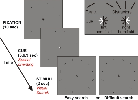
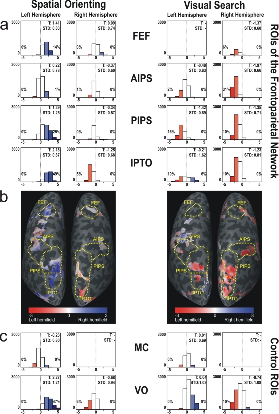
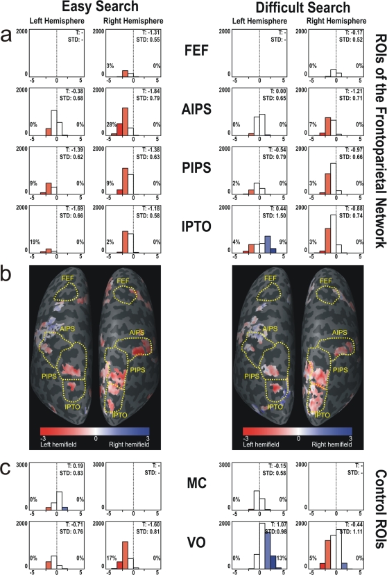
To what extent are the left and right visual hemifields spatially coded in the dorsal frontoparietal attention network? In many experiments with neglect patients, the left hemisphere shows a contralateral hemifield preference, whereas the right hemisphere represents both hemifields. This pattern of spatial coding is often used to explain the right-hemispheric dominance of lesions causing hemispatial neglect. However, pathophysiological mechanisms of hemispatial neglect are controversial because recent experiments on healthy subjects produced conflicting results regarding the spatial coding of visual hemifields. We used an fMRI paradigm that allowed us to distinguish two attentional subprocesses during a visual search task. Either within the left or right hemifield subjects first attended to stationary locations (spatial orienting) and then shifted their attentional focus to search for a target line. Dynamic changes in spatial coding of the left and right hemifields were observed within subregions of the dorsal front-parietal network: During stationary spatial orienting, we found the well-known spatial pattern described above, with a bilateral hemifield representation in the right hemisphere and a contralateral preference in the left hemisphere. However, during search, the right hemisphere had a contralateral preference and the left hemisphere equally represented both hemifields. This finding leads to novel perspectives regarding models of visuospatial attention and hemispatial neglect.
Conflict of interest statement
Figures
Similar articles
-
Influences of Long-Term Memory-Guided Attention and Stimulus-Guided Attention on Visuospatial Representations within Human Intraparietal Sulcus.J Neurosci. 2015 Aug 12;35(32):11358-63. doi: 10.1523/JNEUROSCI.1055-15.2015. J Neurosci. 2015. PMID: 26269642 Free PMC article.
-
Disorders of visuospatial orientation in the frontal plane in patients with visual neglect following right or left parietal lesions.Exp Brain Res. 1998 Sep;122(1):108-20. doi: 10.1007/s002210050497. Exp Brain Res. 1998. PMID: 9772118 Clinical Trial.
-
Acute visual neglect and extinction: distinct functional state of the visuospatial attention system.Brain. 2011 Nov;134(Pt 11):3310-25. doi: 10.1093/brain/awr220. Epub 2011 Sep 23. Brain. 2011. PMID: 21948940
-
The hybrid model of attentional control: New insights into hemispheric asymmetries inferred from TMS research.Neuropsychologia. 2015 Jul;74:21-9. doi: 10.1016/j.neuropsychologia.2014.11.023. Epub 2014 Nov 20. Neuropsychologia. 2015. PMID: 25451041 Review.
-
Spatial attention and neglect: parietal, frontal and cingulate contributions to the mental representation and attentional targeting of salient extrapersonal events.Philos Trans R Soc Lond B Biol Sci. 1999 Jul 29;354(1387):1325-46. doi: 10.1098/rstb.1999.0482. Philos Trans R Soc Lond B Biol Sci. 1999. PMID: 10466154 Free PMC article. Review.
Cited by
-
Spatial distribution of attention and inter-hemispheric competition.Cogn Process. 2015 Nov;16(4):417-25. doi: 10.1007/s10339-015-0734-5. Epub 2015 Aug 20. Cogn Process. 2015. PMID: 26289477
-
Spectral parameters modulation and source localization of blink-related alpha and low-beta oscillations differentiate minimally conscious state from vegetative state/unresponsive wakefulness syndrome.PLoS One. 2014 Mar 27;9(3):e93252. doi: 10.1371/journal.pone.0093252. eCollection 2014. PLoS One. 2014. PMID: 24676098 Free PMC article.
-
Dynamic upper and lower visual field preferences within the human dorsal frontoparietal attention network.Hum Brain Mapp. 2011 Jul;32(7):1036-49. doi: 10.1002/hbm.21087. Epub 2010 Jul 27. Hum Brain Mapp. 2011. PMID: 20665723 Free PMC article.
-
Top-down versus bottom-up attention differentially modulate frontal-parietal connectivity.Hum Brain Mapp. 2020 Mar;41(4):928-942. doi: 10.1002/hbm.24850. Epub 2019 Nov 6. Hum Brain Mapp. 2020. PMID: 31692192 Free PMC article.
References
-
- Danckert JA, Goodale MA. Ups and Downs in the Visual Control of Actions. In: Johnson-Frey SH, editor. Taking Action: Cognitive Neuroscience Perspectives onIntentional Actions. Cambridge, MA: MIT Press; 2003. pp. 29–64.
-
- Niemeier M, Goltz HC, Kuchinad A, Tweed DB, Vilis T. A contralateral preference in the lateral occipital area: sensory and attentional mechanisms. Cereb Cortex. 2005;15:325–331. - PubMed
-
- Corbetta M, Shulman GL. Control of goal-directed and stimulus-driven attention in the brain. Nat Rev Neurosci. 2002;3:201–215. - PubMed
Publication types
MeSH terms
LinkOut - more resources
Full Text Sources
