. 2007 Sep 19;3(9):e3242.
doi: 10.1371/journal.pone.0003242.

The 5' leader of the mRNA encoding the mouse neurotrophin receptor TrkB contains two internal ribosomal entry sites that are differentially regulated

Affiliations

The 5' leader of the mRNA encoding the mouse neurotrophin receptor TrkB contains two internal ribosomal entry sites that are differentially regulated

Stephanie L Timmerman et al. PLoS One. .

Abstract

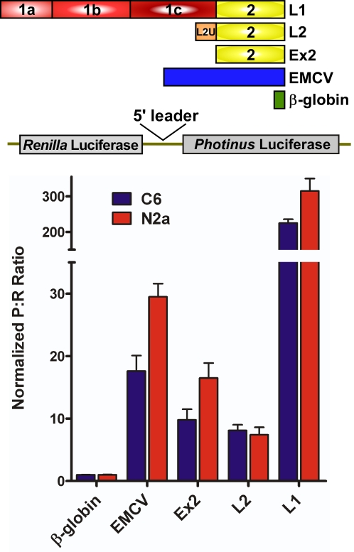
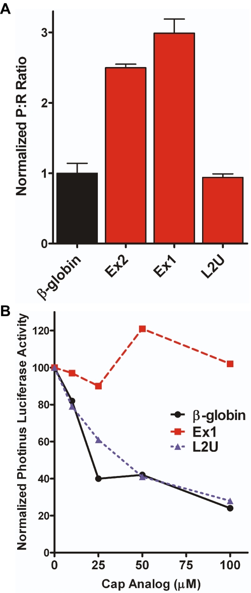
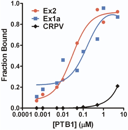
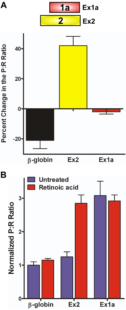
A single internal ribosomal entry site (IRES) in conjunction with IRES transactivating factors (ITAFs) is sufficient to recruit the translational machinery to a eukaryotic mRNA independent of the cap structure. However, we demonstrate that the mouse TrkB mRNA contains two independent IRESes. The mouse TrkB mRNA consists of one of two 5' leaders (1428 nt and 448 nt), both of which include the common 3' exon (Ex2, 344 nt). Dicistronic RNA transfections and in vitro translation of monocistronic RNA demonstrated that both full-length 5' leaders, as well as Ex2, exhibit IRES activity indicating the IRES is located within Ex2. Additional analysis of the upstream sequences demonstrated that the first 260 nt of exon 1 (Ex1a) also contains an IRES. Dicistronic RNA transfections into SH-SY5Y cells showed the Ex1a IRES is constitutively active. However, the Ex2 IRES is only active in response to retinoic acid induced neural differentiation, a state which correlates with the synthesis of the ITAF polypyrimidine tract binding protein (PTB1). Correspondingly, addition or knock-down of PTB1 altered Ex2, but not Ex1a IRES activity in vitro and ex vivo, respectively. These results demonstrate that the two functionally independent IRESes within the mouse TrkB 5' leader are differentially regulated, in part by PTB1.


Conflict of interest statement

Competing Interests: The authors have declared that no competing interests exist.

Figures

Figure 1
Figure 1. The mouse TrkB 5′ leaders.
(A) Schematic representation of the gene structure of the mouse TrkB 5′ leader and variations used for luciferase assays (B) as well as the two controls, the β-globin 5′ leader and EMCV IRES. The boxes represent exons, lines represent introns, and the blue arrows indicate the transcriptional start site of the two promoters.
Figure 2
Figure 2. The mouse TrkB 5′ leaders exhibit an increased Photinus to Renilla luciferase (P∶R) ratio.
Dicistronic luciferase constructs containing the β-globin, EMCV, and TrkB 5′ leaders were transfected into the C6 and N2a neural cell lines. The P∶R ratio from each construct was normalized to that obtained from the β-globin construct, whose P∶R ratio was set to one.
Figure 3
Figure 3. The mouse TrkB 5′ leaders exhibit cryptic promoter activity.
Promoterless dicistronic luciferase constructs containing the β-globin or the TrkB 5′ leaders were transfected into C6 cells. The P∶R ratio was normalized to that obtained from construct containing the β-globin 5′ leader.
Figure 4
Figure 4. The mouse TrkB 5′ leaders exhibit IRES activity when expressed in dicistronic RNA constructs.
Dicistronic luciferase mRNA containing the β-globin and the TrkB 5′ leaders were transfected into C6 cells. The P∶R ratio for each construct was normalized to that of the negative control, β-globin.
Figure 5
Figure 5. The mouse TrkB 5′ leaders are able to initiate translation when cap-dependent translation is inhibited.
A) A dual monocistronic construct containing the pGL3 multiple cloning site upstream of the Renilla luciferase and the pGL3 multiple cloning site, EMCV, or the TrkB 5′ leaders upstream of the Photinus luciferase gene was co-transfected into C6 cells with a construct encoding for a hypophosphorylated 4EBP construct or a null vector. The level of Photinus luciferase activity from each mRNA, when co-transfected with the null vector, was normalized to 100 percent. The Renilla and Photinus luciferase activity obtained in cells co-transfected with hypophosphorylated 4EBP is represented as a percentage of the activity obtained in cells co-transfected with the null plasmid. B) Monocistronic Photinus luciferase mRNA containing the β-globin or TrkB 5′ leaders was in vitro translated in rabbit reticulocyte lysate in the presence of increasing concentrations of cap analog. Photinus luciferase activity for each mRNA in the absence of cap analog was set to 100 percent.
Figure 6
Figure 6. The mouse TrkB mRNA contains two independent IRESes.
A) The additional sequence upstream of Exon 2 from both full-length leaders (Ex1 and L2U) and Exon 2 alone (see Fig 1a) were individually inserted into dicistronic RNA vectors and transfected into C6 cells. The resulting P∶R ratios were normalized to the ratio observed from the mRNA containing the β-globin 5′ leader. B) The unique regions of the full-length TrkB 5′ leaders, as well as the β-globin 5′ leader were inserted upstream of a monocistronic Photinus luciferase open reading frame and in vitro translated in the presence of increasing concentrations of cap analog. The initial level of Photinus luciferase activity was set to 100 percent for each mRNA.
Figure 7
Figure 7. The upstream IRES in the mouse TrkB 5′ leader is located within Ex1a.
The exon 1 splice variants (shown in the schematic, see also Fig 1A) were inserted into dicistronic RNA vectors and transfected into C6 cells. The resulting P∶R ratios were normalized to the ratio observed from β-globin 5′ leader.
Figure 8
Figure 8. The mouse TrkB 5′ leaders bind PTB1 protein.
Purified PTB1 was added in increasing amounts to radiolabeled RNA containing Ex1a, Ex2, or the negative control CrPV IRES. A Langmuir plot was created using the calculated fraction bound. Disassociation constants of 85 nM and 46 nM were determined for the PTB1 interaction with Ex1a and Ex2, respectively.
Figure 9
Figure 9. PTB increases IRES activity from the Ex2 IRES, but does not affect Ex1a IRES activity.
A) Dicistronic luciferase mRNA containing the Ex1a, Ex2, or β-globin 5′ leader was in vitro translated in RRL that was either untreated or supplemented with 0.4 mg of PTB1. The change in the P∶R ratio for the samples when in the presence of PTB1 relative to the untreated sample are shown. B) The two mouse TrkB IRESes demonstrate differential regulation. SH-SY5Y cells treated with 2 mM retinoic acid or with DMSO (mock) for four days were transfected with dicistronic mRNA containing the β-globin, Ex2, or Ex1a 5′ leaders. The P∶R ratios were normalized to that from the mRNA containing the β-globin 5′ leader.
Figure 10
Figure 10. PTB expression is required for IRES activity mediated by Ex2.
A) Western blot analysis of PTB and GAPDH (as a loading control) from lysates obtained from differentiated and undifferentiated SH-SY5Y cells transfected with siRNA directed against PTB or mock transfected for 72 hours. B) Dicistronic RNA containing the two mouse TrkB IRESes were transfected into differentiated SH-SY5Y cells depleted of PTB1 by siRNA. The P∶R ratios were normalized to that from the mRNA containing the β-globin 5′ leader in control undifferentiated SH-SY5Y cells.

References

    1. Kozak M. How do eucaryotic ribosomes select initiation regions in messenger RNA? Cell. 1978;15:1109–1123. - PubMed
    1. Komar AA, Hatzoglou M. Internal ribosome entry sites in cellular mRNAs: mystery of their existence. J Biol Chem. 2005;280:23425–23428. - PubMed
    1. Sonenberg N, Skup D, Trachsel H, Millward S. In vitro translation in reovirus- and poliovirus-infected cell extracts. Effects of anti-cap binding protein monoclonal antibody. J Biol Chem. 1981;256:4138–4141. - PubMed
    1. Belsham GJ, Sonenberg N. Picornavirus RNA translation: roles for cellular proteins. Trends Microbiol. 2000;8:330–335. - PubMed
    1. Mitchell SA, Spriggs KA, Coldwell MJ, Jackson RJ, Willis AE. The Apaf-1 internal ribosome entry segment attains the correct structural conformation for function via interactions with PTB and unr. Mol Cell. 2003;11:757–771. - PubMed

Publication types