

Cryopreservation of composite tissue transplants
- PMID: 18780115
- PMCID: PMC2528968
- DOI: 10.1007/s11552-007-9062-2
Cryopreservation of composite tissue transplants
Abstract
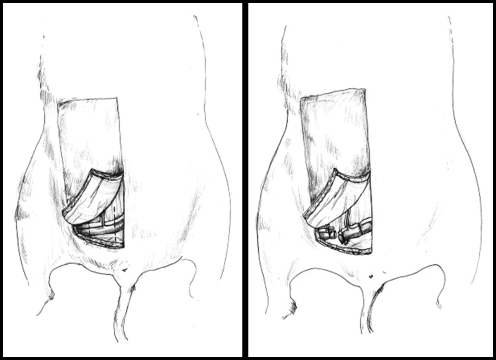
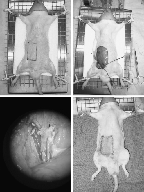
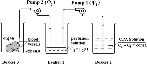
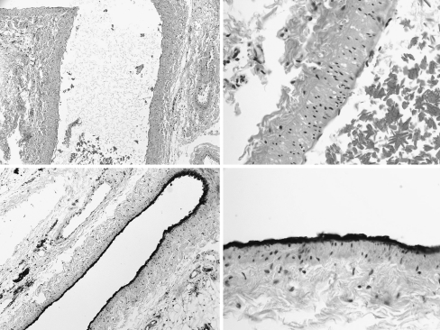
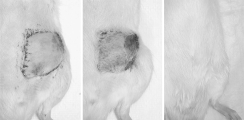
Composite tissue allotransplantation holds great promise for upper extremity reconstruction but is limited by donor part availability. Cryopreservation may increase the availability of donor parts and even reduce antigenicity. The purpose of the study was to evaluate the viability of cryopreserved composite tissues and to demonstrate the feasibility of microvascular isotransplantation of cryopreserved composite flaps. Twenty epigastric flaps were harvested from Lewis rats. Ten flaps were analyzed fresh. Ten flaps were perfused with dimethyl sulfoxide (DMSO)/trehelose cryoprotectant agent (CPA), frozen by controlled cooling to -140 degrees C, and stored for 2 weeks. Flaps were evaluated by factor VIII endothelial staining and MTT tetrazolium salt assay. For the in vivo phase, 30 flaps were harvested. Ten were transplanted fresh to isogenetic recipient animals, ten were perfused with CPA and transplanted, and ten were cryopreserved for 2 weeks, thawed, and transplanted. All cryopreserved samples displayed intact vascular endothelia on factor VIII staining. On MTT analysis, the epithelial viability index for the cryopreserved samples was not significantly different from fresh controls (p = 0.12). All freshly transplanted flaps (10/10) were viable at 60 days. Nine of ten flaps in the perfused/transplanted group were viable at 60 days. Survival of cryopreserved/transplanted flaps ranged from 5 to 60 days. The skin and vascular endothelial components of composite tissue flaps appear to retain their viability after cryopreservation. The in vivo studies demonstrate that the long-term survival of cryopreserved composite tissue transplants is feasible and support an indirect injury, rather than direct injury from freezing or cryoprotectant agents, as the mechanism of flap loss.
Figures
Similar articles
-
Cryopreservation of composite tissues and transplantation: preliminary studies.Cryobiology. 2007 Dec;55(3):295-304. doi: 10.1016/j.cryobiol.2007.08.013. Epub 2007 Sep 18. Cryobiology. 2007. PMID: 17963742
-
Cryopreservation of composite tissue flaps: experimental studies in the rat.Ann Plast Surg. 2007 Jun;58(6):656-60. doi: 10.1097/01.sap.0000245114.96202.62. Ann Plast Surg. 2007. PMID: 17522490
-
Cryopreservation of osteochondral allografts: dimethyl sulfoxide promotes angiogenesis and immune tolerance in mice.J Bone Joint Surg Am. 2002 Aug;84(8):1420-9. J Bone Joint Surg Am. 2002. PMID: 12177273
-
Cryopreserved primary hepatocytes as a constantly available in vitro model for the evaluation of human and animal drug metabolism and enzyme induction.Drug Metab Rev. 2000 Feb;32(1):81-118. doi: 10.1081/dmr-100100564. Drug Metab Rev. 2000. PMID: 10711408 Review.
-
Review of in vivo studies of dimethyl sulfoxide cryopreserved platelets.Transfus Med Rev. 2014 Oct;28(4):212-25. doi: 10.1016/j.tmrv.2014.09.001. Epub 2014 Sep 21. Transfus Med Rev. 2014. PMID: 25439164 Review.
Cited by
-
Pharmacodynamic correlations using fresh and cryopreserved tissue following use of vaginal rings containing dapivirine and/or maraviroc in a randomized, placebo controlled trial.Medicine (Baltimore). 2016 Jul;95(28):e4174. doi: 10.1097/MD.0000000000004174. Medicine (Baltimore). 2016. PMID: 27428211 Free PMC article. Clinical Trial.
-
Cryopreservation and replantation of amputated rat hind limbs.Eur J Med Res. 2014 May 23;19(1):28. doi: 10.1186/2047-783X-19-28. Eur J Med Res. 2014. PMID: 24886622 Free PMC article.
-
Preservation solutions for attenuation of ischemia-reperfusion injury in vascularized composite allotransplantation.SAGE Open Med. 2021 Jul 21;9:20503121211034924. doi: 10.1177/20503121211034924. eCollection 2021. SAGE Open Med. 2021. PMID: 34367640 Free PMC article.
-
Improving the ischemia-reperfusion injury in vascularized composite allotransplantation: Clinical experience and experimental implications.Front Immunol. 2022 Sep 16;13:998952. doi: 10.3389/fimmu.2022.998952. eCollection 2022. Front Immunol. 2022. PMID: 36189311 Free PMC article. Review.
-
Tissue conservation for transplantation.Innov Surg Sci. 2017 Aug 8;2(4):171-187. doi: 10.1515/iss-2017-0010. eCollection 2017 Dec. Innov Surg Sci. 2017. PMID: 31579751 Free PMC article. Review.
References
-
- {'text': '', 'ref_index': 1, 'ids': [{'type': 'DOI', 'value': '10.1097/01.TP.0000100398.39169.5B', 'is_inner': False, 'url': 'https://doi.org/10.1097/01.tp.0000100398.39169.5b'}, {'type': 'PubMed', 'value': '14688529', 'is_inner': True, 'url': 'https://pubmed.ncbi.nlm.nih.gov/14688529/'}]}
- Akst LM, Siemionow M, Dan O, Izycki D, Strome M. Induction of tolerance in a rat model of laryngeal transplantation. Transplantation 2003;76:1763–70. - PubMed
-
- {'text': '', 'ref_index': 1, 'ids': [{'type': 'DOI', 'value': '10.1023/A:1021846703301', 'is_inner': False, 'url': 'https://doi.org/10.1023/a:1021846703301'}, {'type': 'PubMed', 'value': '15256893', 'is_inner': True, 'url': 'https://pubmed.ncbi.nlm.nih.gov/15256893/'}]}
- Alotto D, Ariotti S, Graziano S, Verrua R, Stella M, Magliacani G, et al. The role of quality control in a skin bank: tissue viability determination. Cell and Tissue Banking 2002;3:3–10. - PubMed
-
- {'text': '', 'ref_index': 1, 'ids': [{'type': 'DOI', 'value': '10.1111/j.1600-6143.2005.01144.x', 'is_inner': False, 'url': 'https://doi.org/10.1111/j.1600-6143.2005.01144.x'}, {'type': 'PubMed', 'value': '16433752', 'is_inner': True, 'url': 'https://pubmed.ncbi.nlm.nih.gov/16433752/'}]}
- Birchall MA, Lorenz RR, Berke GS, Genden EM, Haughey BH, Siemionow M, et al. Laryngeal transplantation in 2005: a review. Am J Transplant 2006;6:20–6. - PubMed
-
- {'text': '', 'ref_index': 1, 'ids': [{'type': 'DOI', 'value': '10.1016/j.burns.2003.01.001', 'is_inner': False, 'url': 'https://doi.org/10.1016/j.burns.2003.01.001'}, {'type': 'PubMed', 'value': '14636749', 'is_inner': True, 'url': 'https://pubmed.ncbi.nlm.nih.gov/14636749/'}]}
- Castagnoli C, Alotto D, Cambieri I, Casimiri R, Aluffi M, Stella M, et al. Evaluation of donor skin viability: fresh and cryopreserved skin using tetrazolium salt assay. Burns 2003;29:759–67. - PubMed
-
- {'text': '', 'ref_index': 1, 'ids': [{'type': 'DOI', 'value': '10.1016/S0006-8993(96)01166-3', 'is_inner': False, 'url': 'https://doi.org/10.1016/s0006-8993(96)01166-3'}, {'type': 'PubMed', 'value': '9138719', 'is_inner': True, 'url': 'https://pubmed.ncbi.nlm.nih.gov/9138719/'}]}
- Dawson DA, Ruetzler CA, Hallenbeck JM. Temporal impairment of microcirculatory perfusion following focal cerebral ischemia in the spontaneously hypertensive rat. Brain Res 1997;28:200–8. - PubMed
LinkOut - more resources
Full Text Sources
