

Case Reports
doi: 10.1007/s11999-008-0484-0.
Epub 2008 Sep 9.
Orthopaedic . radiology . pathology conference: left hip pain in a 62-year-old man
Affiliations
- PMID: 18780134
- PMCID: PMC2690732
- DOI: 10.1007/s11999-008-0484-0
Item in Clipboard
Case Reports
Orthopaedic . radiology . pathology conference: left hip pain in a 62-year-old man
Clin Orthop Relat Res.
2009 Jul.
No abstract available
Figures
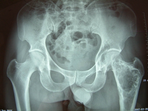
An anteroposterior view of pelvis shows irregularly expansive lytic bone destruction with surrounding sclerosis and endosteal scalloping in the metaphysis of the left femur and femoral head.
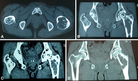
An (A) axial scan of the pelvis shows erosion of the medial cortex with marked thinning and destruction of the cortex posteromedially and thickening anteriorly of the left femur. (B) A coronal reconstruction CT scan of the bony window shows a large osteolytic lesion with spotty calcifications and mixed density in the metaphysis of the left femur and femoral head with erosion of its medial cortex. These coronal reconstruction CT scans of the soft tissue window show (C) some swelling of the soft tissue and (D) an obvious breach of the medial contex of the left femur.
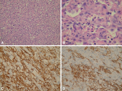
Photomicrographs show histologic sections of the lesion. (A) Solid nests or large sheets of spindle or epithelioid cells are interspersed between and around preexisting vessels with flat endothelium (Stain, hematoxylin and eosin; original magnification, ×40). (B) Tumor cells are large and pleomorphic and show a moderately abundant eosinophilic cytoplasm and a round-to-oval nucleus with one or two prominent nucleoli. Mitotic figures were numerous and frequently abnormal (Stain, hematoxylin and eosin; original magnification, ×400). (C) Immunohistochemical analysis revealed strong staining of tumor cells for CD31 (Original magnification, ×200). (D) Immunostaining for CD34 was partially positive (Original magnification, ×200).
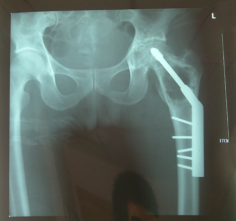
A postoperative anteroposterior view shows the left hip after curettage was performed and a dynamic hip screw was inserted in the intertrochanteric region of the left femur.
Similar articles
-
Angiosarcoma of the chest wall in a patient with fibrous dysplasia.Eur J Cardiothorac Surg. 2002 Oct;22(4):654-5. doi: 10.1016/s1010-7940(02)00399-8. Eur J Cardiothorac Surg. 2002. PMID: 12297197
-
Hip pain in a 21-year-old man.Clin Orthop Relat Res. 1995 Feb;(311):276-7, 287-8. Clin Orthop Relat Res. 1995. PMID: 7634586 No abstract available.
-
Proximal Femur Chondrosarcoma Misdiagnosed as Hip Arthritis: A Case Report.JBJS Case Connect. 2020 Jan-Mar;10(1):e0324. doi: 10.2106/JBJS.CC.19.00324. JBJS Case Connect. 2020. PMID: 32224655
-
Intracortical osteosarcoma.Skeletal Radiol. 1998 Apr;27(4):228-32. doi: 10.1007/s002560050372. Skeletal Radiol. 1998. PMID: 9592909 Review.
-
Metal-associated angiosarcoma of bone: report of two cases and review of the literature.Clin Orthop Relat Res. 2002 Mar;(396):206-14. doi: 10.1097/00003086-200203000-00031. Clin Orthop Relat Res. 2002. PMID: 11859245 Review.
References
-
- {'text': '', 'ref_index': 1, 'ids': [{'type': 'DOI', 'value': '10.1007/BF00266872', 'is_inner': False, 'url': 'https://doi.org/10.1007/bf00266872'}, {'type': 'PubMed', 'value': '4040502', 'is_inner': True, 'url': 'https://pubmed.ncbi.nlm.nih.gov/4040502/'}]}
- Adler CP, Reichelt A. Haemangiosarcoma of bone. Int Orthop. 1985;8:273−279. - PubMed
-
- {'text': '', 'ref_index': 1, 'ids': [{'type': 'PMC', 'value': 'PMC8337554', 'is_inner': False, 'url': 'https://pmc.ncbi.nlm.nih.gov/articles/PMC8337554/'}, {'type': 'PubMed', 'value': '8933884', 'is_inner': True, 'url': 'https://pubmed.ncbi.nlm.nih.gov/8933884/'}]}
- Bourekas EC, Cohen ML, Kamen CS, Tarr RW, Lanzieri CF, Lewin JS. Malignant hemangioendothelioma (angiosarcoma) of the skull: plain film, CT, and MR appearance. AJNR Am J Neuroradiol. 1996;17:1946–1948. - PMC - PubMed
-
- {'text': '', 'ref_index': 1, 'ids': [{'type': 'PubMed', 'value': '9728613', 'is_inner': True, 'url': 'https://pubmed.ncbi.nlm.nih.gov/9728613/'}]}
- De Young BR, Frierson HF Jr, Ly MN, Smith D, Swanson PE. CD31 immunoreactivity in carcinomas and mesotheliomas. Am J Clin Pathol. 1998;110:374–377. - PubMed
-
- {'text': '', 'ref_index': 1, 'ids': [{'type': 'DOI', 'value': '10.1002/1097-0142(19950101)75:1+<203::AID-CNCR2820751308>3.0.CO;2-V', 'is_inner': False, 'url': 'https://doi.org/10.1002/1097-0142(19950101)75:1+<203::aid-cncr2820751308>3.0.co;2-v'}, {'type': 'PubMed', 'value': '8000997', 'is_inner': True, 'url': 'https://pubmed.ncbi.nlm.nih.gov/8000997/'}]}
- Dorfman HD, Czerniak B. Bone cancers. Cancer. 1995;75(1 suppl):203–210. - PubMed
-
- {'text': '', 'ref_index': 1, 'ids': [{'type': 'PubMed', 'value': '8311392', 'is_inner': True, 'url': 'https://pubmed.ncbi.nlm.nih.gov/8311392/'}]}
- Goldstein WS, Bowen BC, Balkany T. Malignant hemangioendothelioma of the temporal bone masquerading as glomus tympanicum. Ann Otol Rhinol Laryngol. 1994;103:156–159. - PubMed
Publication types
MeSH terms
LinkOut - more resources
Full Text Sources
