

Monocyte/macrophage trafficking in acquired immunodeficiency syndrome encephalitis: lessons from human and nonhuman primate studies
- PMID: 18780233
- PMCID: PMC2728912
- DOI: 10.1080/13550280802132857
Monocyte/macrophage trafficking in acquired immunodeficiency syndrome encephalitis: lessons from human and nonhuman primate studies
Abstract
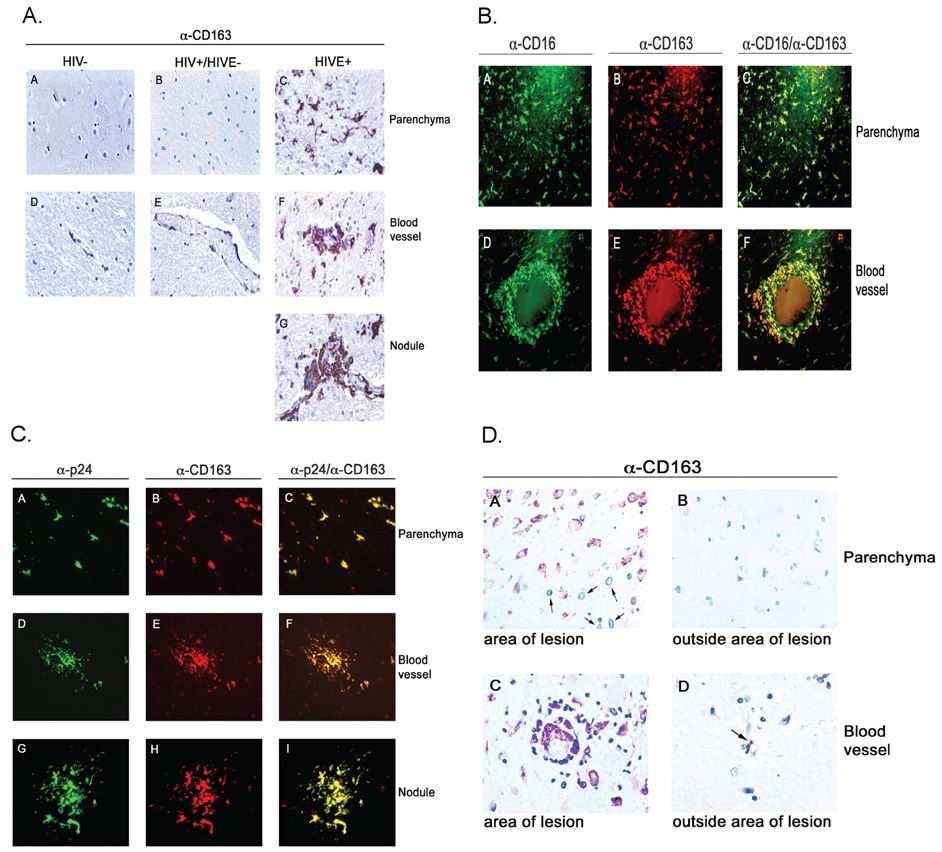
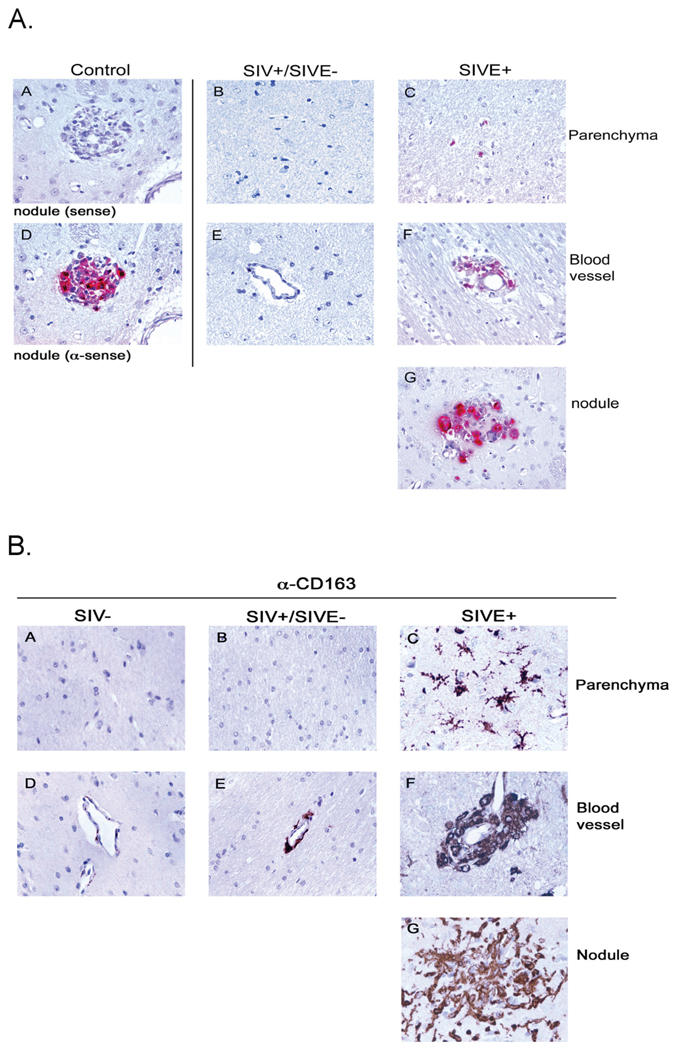
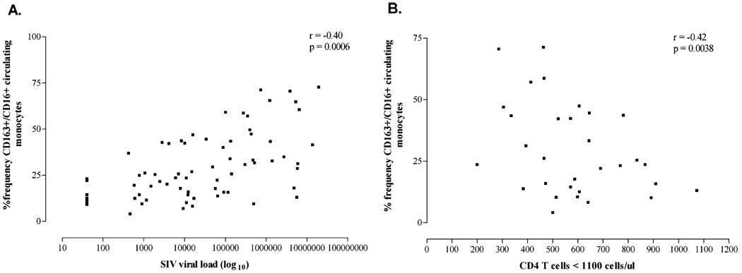
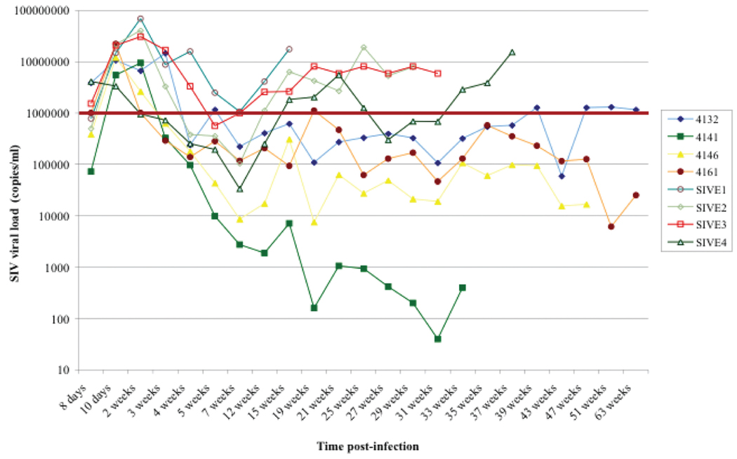
Here the authors discuss evidence in human and animal models supporting two opposing views regarding the pathogenesis of human immunodeficiency virus (HIV) in the central nervous system (CNS): (1) HIV infection in the CNS is a compartmentalized infection, with the virus-infected macrophages entering the CNS early, infecting resident microglia and astrocytes, and achieving a state of latency with evolution toward a fulminant CNS infection late in the course of disease; or alternatively, (2) events in the periphery lead to altered monocyte/macrophage (MPhi) homeostasis, with increased CNS invasion of infected and/or uninfected MPhis. Here the authors have reevaluated evidence presented in the favor of the latter model, with a discussion of phenotypic characteristics distinguishing normal resident microglia with those accumulating in HIV encephalitis (HIVE). CD163 is normally expressed by perivascular MPhi s but not resident microglia in normal CNS of humans and rhesus macaques. In agreement with other studies, the authors demonstrate expression of CD163 by brain MPhi s in HIVE and simian immunodeficiency virus encephalitis (SIVE). CNS tissues from HIV-sero positive individuals with HIVE or HIV-associated progressive multifocal leukoencephalopathy (PML) were also examined. In HIVE, the authors further demonstrate colocalization of CD163 and CD16 (Fcgamma III recptor) gene expression, the latter marker associated with HIV infection of monocyte in vivo and permissivity of infection. Indeed, CD163(+) MPhis and microglia are often productively infected in HIVE CNS. In SIV infected rhesus macaques, CD163(+) cells accumulate perivascularly, within nodular lesions and the parenchyma in animals with encephalitis. Likewise, parenchymal microglia and perivascular MPhi s are CD163(+) in HIVE. In contrast to HIVE, CD163(+)perivascular and parenchymal MPhi s in HIV-associated PML were only associated with areas of demyelinating lesions. Interestingly, SIV-infected rhesus macaques whose viral burden was predominantly at 1 x 10(6) copies/ml or greater developed encephalitis. To further investigate the relationship between CD163(+)/CD16(+) MPhis/microglia in the CNS and altered homeostasis in the periphery, the authors performed flow-cytometric analyses of peripheral blood mononuclear cells (PBMCs) from SIV-infected rhesus macaques. The results demonstrate an increase in the percent frequency of CD163(+)/CD16(+) monocytes in animals with detectable virus that correlated significantly with increased viral burden and CD4(+) T-cell decline. These results suggest the importance of this monocyte subset in HIV/SIV CNS disease, and also in the immune pathogenesis of lentiviral infection. The authors further discuss the potential role of CD163(+)/CD16(+) monocyte/MPhi subset expansion, altered myeloid homeostasis, and potential consequences for immune polarization and suppression. The results and discussion here suggest new avenues for the development of acquired immunodeficiency syndrome (AIDS) therapeutics and vaccine design.
Figures
Similar articles
-
Simian Immunodeficiency Virus-Infected Memory CD4+ T Cells Infiltrate to the Site of Infected Macrophages in the Neuroparenchyma of a Chronic Macaque Model of Neurological Complications of AIDS.mBio. 2020 Apr 21;11(2):e00602-20. doi: 10.1128/mBio.00602-20. mBio. 2020. PMID: 32317323 Free PMC article.
-
CD163, a marker of perivascular macrophages, is up-regulated by microglia in simian immunodeficiency virus encephalitis after haptoglobin-hemoglobin complex stimulation and is suggestive of breakdown of the blood-brain barrier.Am J Pathol. 2008 Mar;172(3):725-37. doi: 10.2353/ajpath.2008.070848. Epub 2008 Feb 14. Am J Pathol. 2008. PMID: 18276779 Free PMC article.
-
Role for cFMS in maintaining alternative macrophage polarization in SIV infection: implications for HIV neuropathogenesis.J Neuroinflammation. 2015 Mar 25;12:58. doi: 10.1186/s12974-015-0272-1. J Neuroinflammation. 2015. PMID: 25886134 Free PMC article.
-
Brain macrophages harbor latent, infectious simian immunodeficiency virus.AIDS. 2019 Dec 1;33 Suppl 2(Suppl 2):S181-S188. doi: 10.1097/QAD.0000000000002269. AIDS. 2019. PMID: 31789817 Free PMC article. Review.
-
An SIV macaque model of SIV and HAND: the need for adjunctive therapies in HIV that target activated monocytes and macrophages.J Neurovirol. 2018 Apr;24(2):213-219. doi: 10.1007/s13365-018-0616-6. Epub 2018 Feb 12. J Neurovirol. 2018. PMID: 29435829 Review.
Cited by
-
Molecular mechanisms of neuroinvasion by monocytes-macrophages in HIV-1 infection.Retrovirology. 2010 Apr 7;7:30. doi: 10.1186/1742-4690-7-30. Retrovirology. 2010. PMID: 20374632 Free PMC article. Review.
-
Flipping the switches: CD40 and CD45 modulation of microglial activation states in HIV associated dementia (HAD).Mol Neurodegener. 2011 Jan 11;6(1):3. doi: 10.1186/1750-1326-6-3. Mol Neurodegener. 2011. PMID: 21223591 Free PMC article.
-
Cognitive consequences of a sustained monocyte type 1 IFN response in HIV-1 infection.Curr HIV Res. 2014;12(2):77-84. doi: 10.2174/1570162x12666140526113544. Curr HIV Res. 2014. PMID: 24862334 Free PMC article. Review.
-
Simian immunodeficiency virus-infected rhesus macaques with AIDS co-develop cardiovascular pathology and encephalitis.Front Immunol. 2023 Oct 27;14:1240946. doi: 10.3389/fimmu.2023.1240946. eCollection 2023. Front Immunol. 2023. PMID: 37965349 Free PMC article.
-
Update on Central Nervous System Effects of HIV in Adolescents and Young Adults.Curr HIV/AIDS Rep. 2023 Apr;20(2):19-28. doi: 10.1007/s11904-023-00651-3. Epub 2023 Feb 21. Curr HIV/AIDS Rep. 2023. PMID: 36809477 Free PMC article. Review.
References
-
- Buechler C, Ritter M, Orso E, Langmann T, Klucken J, Schmitz G. Regulation of scavenger receptor CD163 expression in human monocytes and macrophages by pro- and antiinflammatory stimuli. J Leukoc Biol. 2000;67:97–103. - PubMed
-
- Caselli RJ, Beach TG, Yaari R, Reiman EM. Alzheimer’s disease a century later. J Clin Psychiatry. 2006;67:1784–1800. - PubMed
-
- Clements JE, Babas T, Mankowski JL, Suryanarayana K, Piatak M, Jr, Tarwater PM, Lifson JD, Zink MC. The central nervous system as a reservoir for simian immunodeficiency virus (SIV): steady-state levels of SIV DNA in brain from acute through asymptomatic infection. J Infect Dis. 2002;186:905–913. - PubMed
-
- Clements JE, Li M, Gama L, Bullock B, Carruth LM, Mankowski JL, Zink MC. The central nervous system is a viral reservoir in simian immunodeficiency virus-infected macaques on combined antiretroviral therapy: a model for human immunodeficiency virus patients on highly active antiretroviral theraby. J Neuro Virol. 2005;11:180–189. - PubMed
Publication types
MeSH terms
Grants and funding
LinkOut - more resources
Full Text Sources
Other Literature Sources
Medical
Research Materials
