. 2008 Oct;291(10):1301-33.
doi: 10.1002/ar.20758.

Architectonic subdivisions of neocortex in the gray squirrel (Sciurus carolinensis)

Affiliations

Architectonic subdivisions of neocortex in the gray squirrel (Sciurus carolinensis)

Peiyan Wong et al. Anat Rec (Hoboken). 2008 Oct.

Abstract

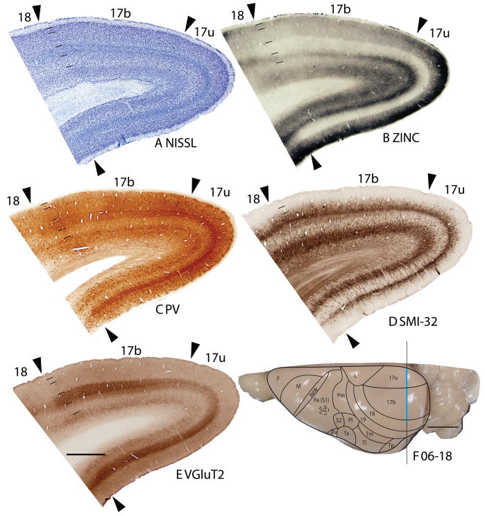
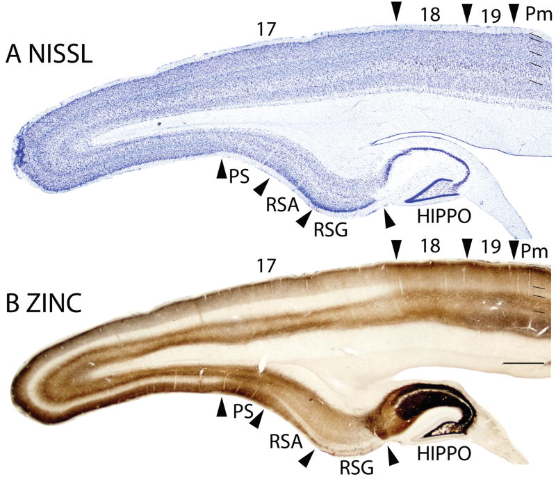
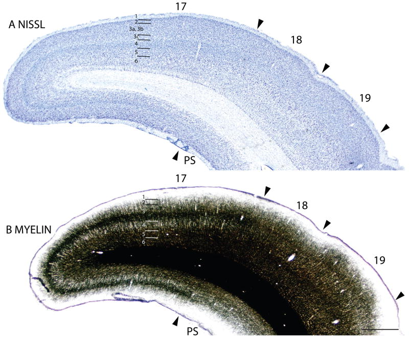
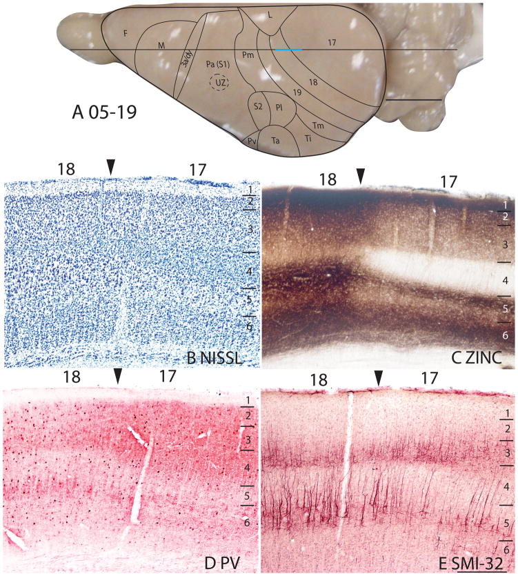
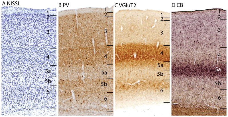
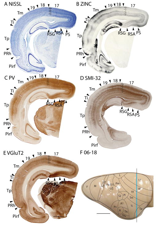
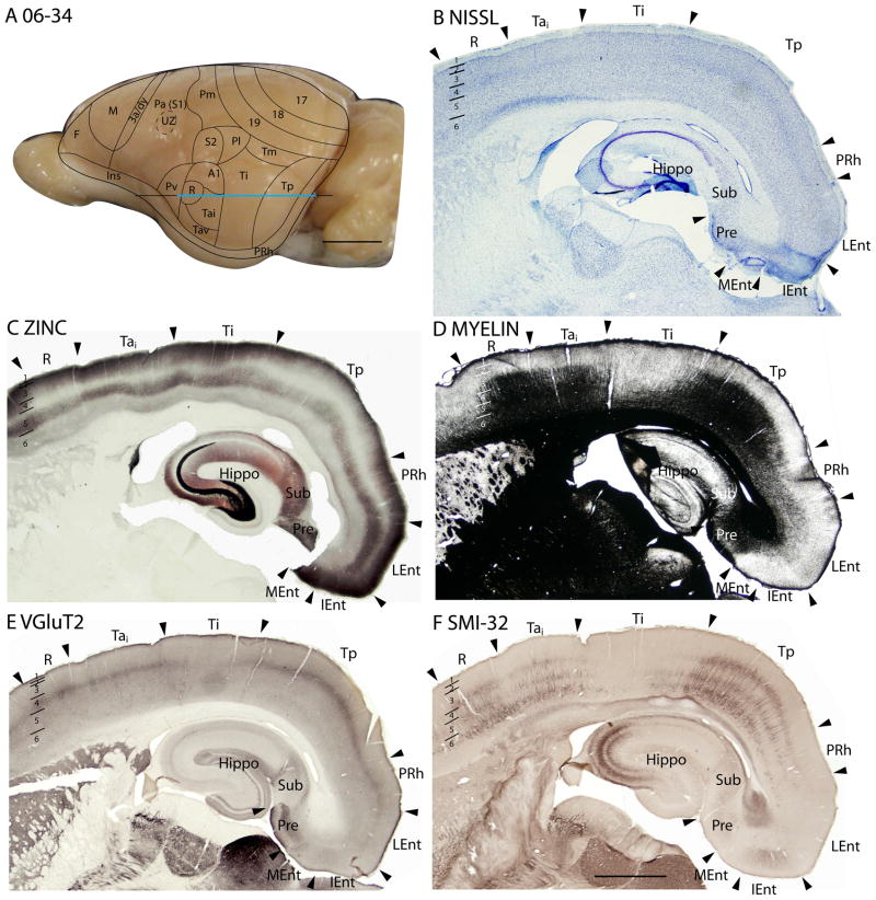
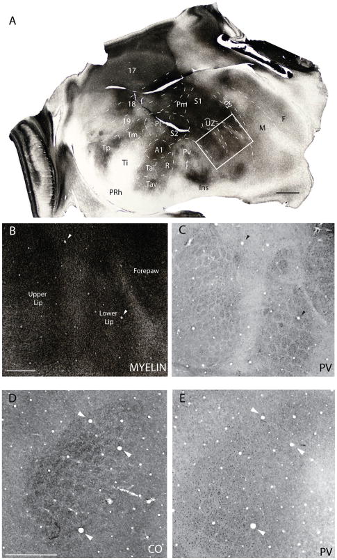
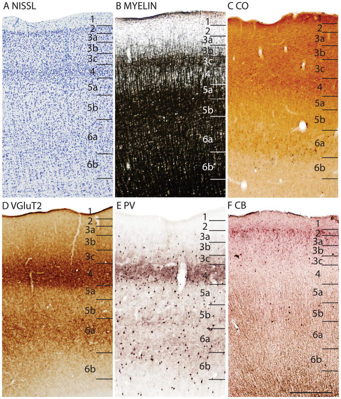
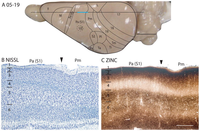
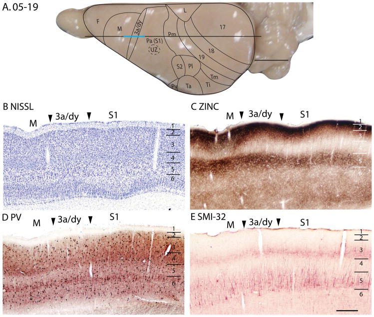
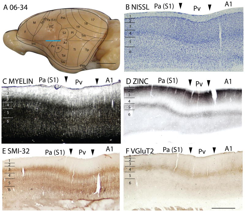
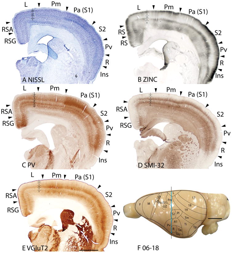
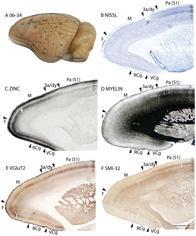
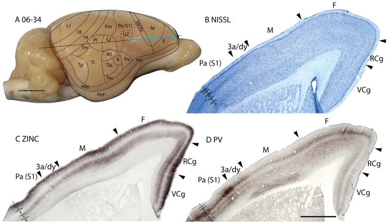
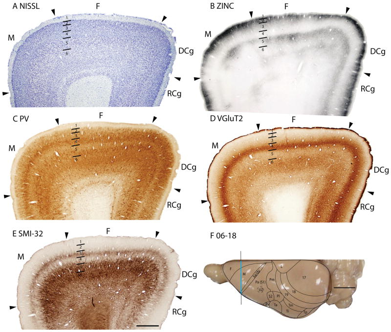
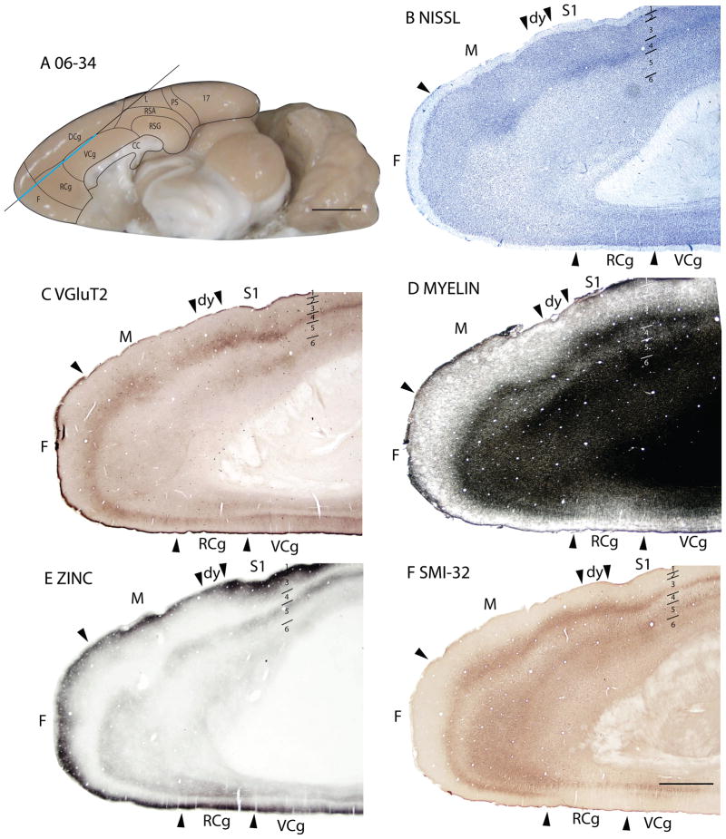
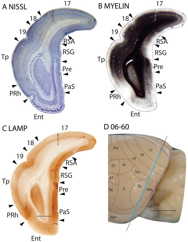
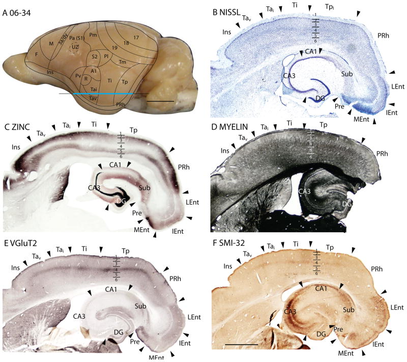
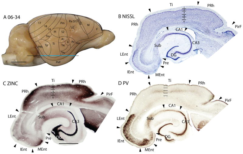
Squirrels are highly visual mammals with an expanded cortical visual system and a number of well-differentiated architectonic fields. To describe and delimit cortical fields, subdivisions of cortex were reconstructed from serial brain sections cut in the coronal, sagittal, or horizontal planes. Architectonic characteristics of cortical areas were visualized after brain sections were processed with immunohistochemical and histochemical procedures for revealing parvalbumin, calbindin, neurofilament protein, vesicle glutamate transporter 2, limbic-associated membrane protein, synaptic zinc, cytochrome oxidase, myelin or Nissl substance. In general, these different procedures revealed similar boundaries between areas, suggesting that functionally relevant borders were being detected. The results allowed a more precise demarcation of previously identified areas as well as the identification of areas that had not been previously described. Primary sensory cortical areas were characterized by sparse zinc staining of layer 4, as thalamocortical terminations lack zinc, as well as by layer 4 terminations rich in parvalbumin and vesicle glutamate transporter 2. Primary areas also expressed higher levels of cytochrome oxidase and myelin. Primary motor cortex was associated with large SMI-32 labeled pyramidal cells in layers 3 and 5. Our proposed organization of cortex in gray squirrels includes both similarities and differences to the proposed of cortex in other rodents such as mice and rats. The presence of a number of well-differentiated cortical areas in squirrels may serve as a guide to the identification of homologous fields in other rodents, as well as a useful guide in further studies of cortical organization and function.


Figures

Figure 1
Figure 1
Architectonic characteristics of visual areas 17 and 18. Coronal sections from occipital cortex were processed for (A) Nissl substance, (B) synaptic zinc, (C) parvalbumin (PV), (D) neurofilaments with the SMI-32 antibody, or (E) the vesicle glutamate transporter 2 (VGluT2). The boundaries of proposed cortical areas are shown on a dorsal view of a squirrel brain in panel F. The vertical line through areas 17 and 18 indicates the locations where sections were taken for panels A–E. The blue line marks the regions shown in these sections. Occipital areas 17, 18 and 19 are adopted from Brodmann (1909). 17u refers to the monocular region, while 17b refers to the binocular region of area 17. Arrowheads mark architectonic boundaries. Short lines under 17/18 arrow heads separate cortical layers 1–6. See table 1 for abbreviations for other areas. The scale bar for brain sections (panel E) = 2mm. The scale bar on the brain (panel F) = 5mm. Sections were from squirrel 06-18.
Figure 2
Figure 2
Architectonic characteristics of visual and adjoining retrosplenial cortex. Brain sections were cut in the parasagittal plane. The sections processed for Nissl substance (A) and zinc (B) were from near the medial wall of the caudal hemisphere. See table 1 for abbreviations. Scale bar = 2mm. Sections were from 05-19. 2. Architectonic characteristics of visual and adjoining retrosplenial cortex. Brain sections were cut in the parasagittal plane. The sections processed for Nissl substance (A) and zinc (B) were from near the medial wall of the caudal hemisphere. See table 1 for abbreviations. Scale bar = 2mm. Sections were from 05-19.
Figure 3
Figure 3
Adjacent coronal brain sections through occipital cortex stained for Nissl substance (A) or myelin (B). Scale bar = 2mm. Sections were from 06-60.
Figure 4
Figure 4
Architectonic characteristics of the bordering region between area 17 and 18. The horizontal line across the dorsal view of the brain in panel A indicates the location of the sagittal sections used in this figure, and the blue line marks the extent of the sections shown in panels B–E. The higher magnification of these panels than in previous figures allows some of the laminar features of area 17 and 18 to be seen more distinctly. The scale bar in panel A = 5mm, panel E = 0.5mm. Squirrel 05-19. The cortical areas depicted on the dorsal view of a squirrel brain in Panel A and similar views in subsequent figures are based on present and previous architectonic and physiological results (see Methods).
Figure 5
Figure 5
The laminar architecture of area 17 at higher magnification. Note how the PV, VGluT2 and CB preparations reveal sublayers. Scare bar = 0.5mm. Sections are in the sagittal plane, from case 05-19.
Figure 6
Figure 6
Architectonic characteristics of subdivisions of occipital and temporal cortex in squirrel 06-18. Borders of cortical layers are marked at the 17/18 boundary. The blue portion of the vertical line across the cortex indicates the location where the coronal brain sections in panels A–E were obtained. Scale bar in panel E = 2mm, panel F = 5mm.
Figure 7
Figure 7
Architectonic characteristics of subdivisions of temporal cortex in squirrel 06-34. Cortical areas are shown on a lateral view of the left caudal hemisphere in panel A. The blue part of the horizontal line across the brain indicates the location of the horizontal brain sections illustrated in panels B–F. Scale bar in panel A = 5mm, panel F = 2mm.
Figure 8
Figure 8
Barrel field of the grey squirrel. A. A myelin stained section cut parallel to the surface of an artificially flattened cerebral hemisphere. Dashed lines show approximate cortical boundaries comparable to the reconstructed dorsal view of the brain in Fig. 1. The boxed region in A is shown in B and C at higher magnification in myelin and PV preparations respectively. D and E are from a separate case and show the barrel field in cytochrome oxidase (CO) and PV preparations respectively. See table 1 for abbreviations. Scale bar for flattened section = 2.0mm, for B and C = 1.0mm, for D and E = 2.0mm.
Figure 9
Figure 9
The laminar architecture of area Pa(S1) at higher magnification. Scale bar = 0.5mm.
Figure 10
Figure 10
Architectonic characteristics of subdivisions of the somatosensory cortex in squirrel 05-19. The horizontal line across the dorsal view of the brain in panel A indicates the location of the sagittal sections used in this figure, and the blue line marks the extent of the sections shown in panels B and C. The extent of each cortical layers 1 to 6 is indicated by the short horizontal lines on panels B–E. The scale bar on the brain (panel A) = 5mm. The scale bar for brain sections (panel E) = 0.5mm.
Figure 11
Figure 11
Architectonic characteristics of subdivisions of the motor and somatosensory cortex in squirrel 05-19. The horizontal line across the dorsal view of the brain in panel A indicates the location of the sagittal sections used in this figure, and the blue line marks the extent of the sections shown in panels B–E. The extent of each cortical layers 1 to 6 is indicated by the short horizontal lines on panels B–E. Scale bar on the brain (panel A) = 5mm. Scale bar for brain sections (panel E) = 0.5mm.
Figure 12
Figure 12
Architectonic characteristics of subdivisions of parietal and primary auditory cortices in squirrel 06-34. Cortical areas are shown on a lateral view of the left hemisphere in panel A. The blue horizontal line across the brain indicates the location of the brain sections illustrated in panels B–F. Short horizontal lines on panels B–F indicate the extent of the 6 cortical layers. Scale bar on the brain (panel A) = 5mm. Scale bar for brain sections (panel F) = 1mm.
Figure 13
Figure 13
Architectonic characteristics of subdivisions of the parietal and limbic cortices in squirrel 06-18. Borders of cortical layers are marked at the limbic (L) area. The blue vertical line across the cortex (panel F) indicates the location where the brain sections in panels A–E were obtained. Short horizontal lines on panels A–E indicate the extent of the 6 cortical layers. Scale bar in panel E = 2mm, panel F = 5mm.
Figure 14
Figure 14
Architectonic characteristics of subdivisions of frontal and cingulate cortices in squirrel 06-34. Cortical areas are shown on a lateral view of the left rostral hemisphere in panel A. The blue horizontal line across the brain indicates the location of the brain sections illustrated in panels B–F. Short horizontal lines on panels B–Fshow the extent of the cortical layers. Scale bar on the brain (panel A) = 5mm. Scale bar for brain sections (panel F) = 2mm.
Figure 15
Figure 15
Architectonic characteristics of subdivisions of frontal and cingulate cortices in squirrel 06-34. Cortical areas are shown on a lateral view of the right rostral hemisphere in panel A. The blue horizontal line across the brain indicates the location of the brain sections illustrated in panels B–D. Short horizontal lines on panels B–D show the extent of the cortical layers. Scale bar on the brain (panel A) = 5mm. Scale bar for brain sections (panel D) = 2mm.
Figure 16
Figure 16
Architectonic characteristics of subdivisions of the frontal cortex in squirrel 06-18. Borders of cortical layers are marked at the frontal (F) area. The blue vertical line across the cortex (panel F) indicates the location where the brain sections in panels A–E were obtained. Scale bar in panel E = 2mm, panel F = 5mm.
Figure 17
Figure 17
Architectonic characteristics of subdivisions of frontal and cingulate cortices in squirrel 06-34. Cortical areas are shown on a medial view of the left hemisphere in panel A. The blue horizontal line across the brain indicates the location of the brain sections illustrated in panels B–F. Short horizontal lines on panels B–F show the extent of the cortical layers. Scale bar on the brain (panel A) = 5mm. Scale bar for brain sections (panel F) = 2mm.
Figure 18
Figure 18
Architectonic characteristics of subdivisions of retrosplenial cortex in squirrel 06-60. Cortical areas are shown on a lateral view of the left caudal hemisphere in panel D. The blue horizontal line across the brain indicates the location of the brain sections illustrated in panels A–C. Short horizontal lines on panels A–C show the extent of the cortical layers. The limbic areas show darker staining in the Limbic Associated Membrane protein (LAMP) compared to other cortical areas, such as area 17 and 18. Scale bar on the brain (panel D) = 5mm. Scale bar for brain sections (panel C) = 2mm.
Figure 19
Figure 19
Architectonic characteristics of subdivisions of temporal and insular cortices in squirrel 06-34. Cortical areas are shown on a medial view of the left hemisphere in panel A. The blue horizontal line across the brain indicates the location of the brain sections illustrated in panels B–F. Short horizontal lines on panels B–F show the extent of the cortical layers. Scale bar on the brain (panel A) = 5mm. Scale bar for brain sections (panel F) = 2mm.
Figure 20
Figure 20
Architectonic characteristics of subdivisions of rhinal cortex in squirrel 06-34. Cortical areas are shown on a lateral view of the right hemisphere in panel A. The blue horizontal line across the brain indicates the location of the brain sections illustrated in panels B–D. Short horizontal lines on panels B–D show the extent of the cortical layers. Scale bar on the brain (panel A) = 5mm. Scale bar for brain sections (panel D) = 2mm.

Similar articles

Cited by

References

    1. Abplanalp P. Some subcortical connections of the visual system in tree shrews and squirrels. Brain Behav Evol. 1970;3(1):155–168. - PubMed
    1. Albus K, Beckmann R. Second and third visual area of the cat: interindividual variability in retinotopic arrangement and cortical location. J Physiol. 1980;299:276–297. - PMC - PubMed
    1. Allman JM, Kaas JH. Representation of the visual field in striate and adjoining cortex of the owl monkey (Aotus trivirgatus) Brain Res. 1971;35(1):89–106. - PubMed
    1. Annese J, Pitiot A, Dinov ID, Toga AW. A myelo-architectonic method for the structural classification of cortical areas. Neuroimage. 2004;21(1):15–26. - PubMed
    1. Avendaño C, Verdu A. Area 3a in the cat. I. A reevaluation of its location and architecture on the basis of Nissl, myelin, acetylcholinesterase, and cytochrome oxidase staining. J Comp Neurol. 1992;321(3):357–72. - PubMed

Publication types

MeSH terms

LinkOut - more resources