. 2008 Oct;180(2):845-56.
doi: 10.1534/genetics.108.092494. Epub 2008 Sep 9.

Secondary mutations correct fitness defects in Toxoplasma gondii with dinitroaniline resistance mutations

Affiliations

Secondary mutations correct fitness defects in Toxoplasma gondii with dinitroaniline resistance mutations

Christopher Ma et al. Genetics. 2008 Oct.

Abstract

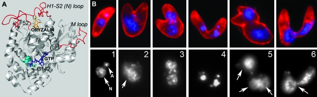
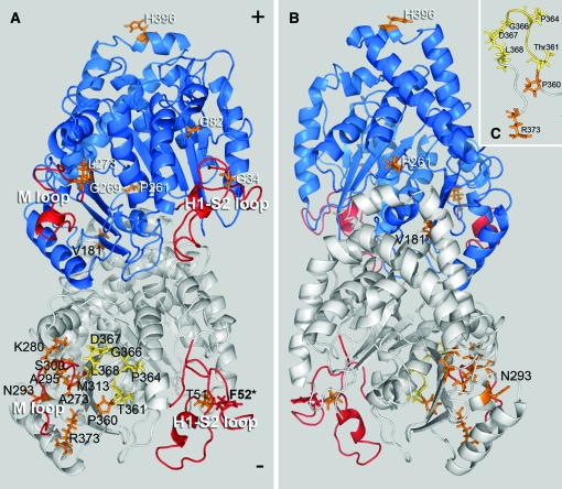
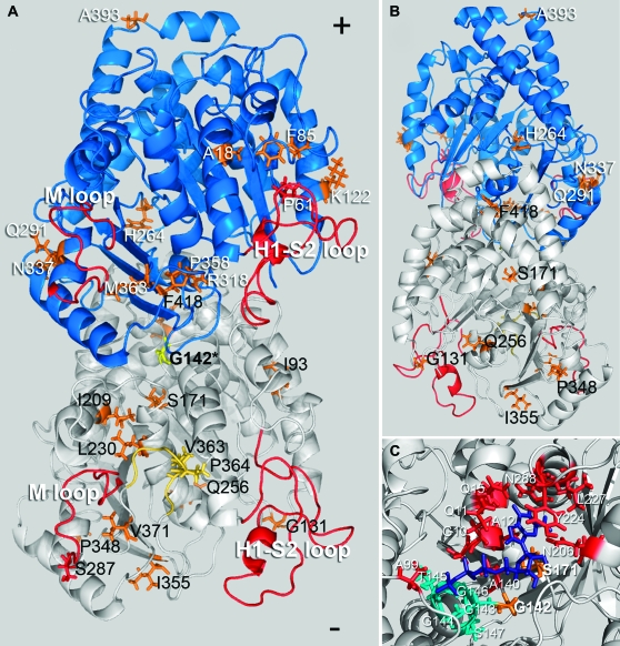
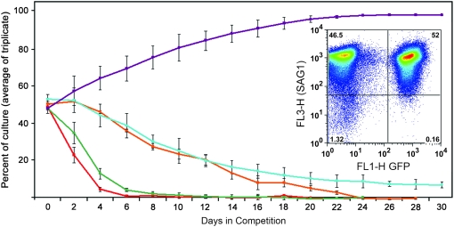
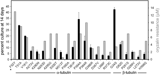
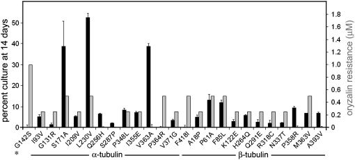
Dinitroanilines (oryzalin, trifluralin, ethafluralin) disrupt microtubules in protozoa but not in vertebrate cells, causing selective death of intracellular Toxoplasma gondii parasites without affecting host cells. Parasites containing alpha1-tubulin point mutations are dinitroaniline resistant but show increased rates of aberrant replication relative to wild-type parasites. T. gondii parasites bearing the F52Y mutation were previously demonstrated to spontaneously acquire two intragenic mutations that decrease both resistance levels and replication defects. Parasites bearing the G142S mutation are largely dependent on oryzalin for viable growth in culture. We isolated 46 T. gondii lines that have suppressed microtubule defects associated with the G142S or the F52Y mutations by acquiring secondary mutations. These compensatory mutations were alpha1-tubulin pseudorevertants or extragenic suppressors (the majority alter the beta1-tubulin gene). Many secondary mutations were located in tubulin domains that suggest that they function by destabilizing microtubules. Most strikingly, we identified seven novel mutations that localize to an eight-amino-acid insert that stabilizes the alpha1-tubulin M loop, including one (P364R) that acts as a compensatory mutation in both F52Y and G142S lines. These lines have reduced dinitroaniline resistance but most perform better than parental lines in competition assays, indicating that there is a trade-off between resistance and replication fitness.


Figures

F<sc>igure</sc> 1.—
Figure 1.—
(A) The F52Y and G142S α-tubulin point mutations confer 13 and 1 μm oryzalin resistance when integrated into wild-type (sensitive) T. gondii parasites. The F52Y mutation (red) is located in the H1-S2 (N) loop (red) of α1-tubulin. The computationally determined dinitroaniline-binding site is below this loop (oryzalin in orange). Interactions between the H1-S2 (N) loop and the M loop of adjacent dimers coordinate lateral associations between protofilaments in the microtubule lattice. The G142S mutation is located in the core of α1-tubulin and the G142 residue represents the initial glycine in the GGGTGSG motif (teal), which is characteristic of tubulins. This motif is part of the GTP-binding site, which is essential for correct folding of the tubulin dimer and interacts with the phosphate portion of GTP (purple). (B) (Top) Immunofluorescence images of extracellular parasites of the G142S line labeled with DG52 (red labels the parasite surface) and DAPI (blue labels the parasite DNA). (Bottom) DNA distribution only. (1) A normal-appearing parasite has a crescent shape and DNA staining of the nucleus (arrow) and apicoplast DNA (arrowhead). (2–6) Parasite “monsters” result from replication defects due to improper coordination of spindle microtubule (nuclear division) and subpellicular microtubule (cytokinesis) functions. Although the extracellular parasites in 3 and 4 are similar to intracellular replicating stages (see diagram in Ma et al. 2007), parasites cannot complete division outside of host cells and their abnormal shape prevents invasion of new host cells. In addition, 2, 5, and 6 illustrate parasites with diffuse (2), improperly segregated (three nuclei) (5), and nonsegregated (6) nuclear staining (compare to 1).
F<sc>igure</sc> 2.—
Figure 2.—
Location of compensatory mutations obtained from the F52Y line mapped on a model of the Toxoplasma tubulin dimer. The α1-subunit is white and the β1-subunit is blue. (A) Most mutations fall between the M and H1-S2 loops (red) facing the inner lumen of the microtubule. The F52Y mutation is located in the H1-S2 loop of α1-tubulin (red) and secondary mutations in α1-tubulin (black text) occur at T51A, V181I, A273V, K280N, N293D, A295G, S300T, M313T, P360A, T361P, P364A, P364R, G366R, D367V, L368F, and R373C (orange/yellow). Mutations in yellow localize to the α-tubulin-specific insert. Extragenic suppressors in the β1-tubulin subunit (white text) occur at G34S, G82D, P261S, G269V, L273V, and H396Q. (B) The β1-tubulin mutation P261S is the only mutation that faces the outer surface of the dimer within the microtubule lattice, while the β-subunit mutation H396Q is located at the dimer–dimer interface within the protofilament. (C) The mutations T361P, P364A, P364R, G366R, D367V, and L368F occur in the α-tubulin-specific insert (yellow) and the mutations P360A and R373C are adjacent to this insert (orange).
F<sc>igure</sc> 3.—
Figure 3.—
Location of compensatory mutations obtained from the G142S line mapped on a model of the Toxoplasma tubulin dimer (the α1-subunit is white and the β1-subunit is blue). The G142S mutation (yellow) is located in the GTP-binding domain of α-tubulin. (A) Most mutations (orange/yellow) are on the surface of the dimer that faces the inner lumen of the microtubule. Secondary mutations in α1-tubulin (black text) occur at I93V, G131R, S171A, I209V, L230V, Q256H, S287P, P348L, I355E, V363A, P364R, V371G, and F418I. Suppressors in the β1-tubulin subunit (white text) occur at residues A18P, P61A, F85L, K122E, H264Q, Q291E, R318C, N337T, P358R, M363V, and A393V. G142S α1-tubulin mutations at V363A and P364R occur in the α-tubulin-specific insert (yellow). (B) The α1-tubulin mutation at Q256 and the β1-tubulin mutation at H264 are the only mutations that face the outer surface of the dimer within the microtubule lattice. The β-subunit mutation A393V is located at the dimer–dimer interface within the protofilament. (C) The primary resistance mutation at G142 is located within the tubulin motif (teal) of the α-tubulin GTP-binding site. The GTP moiety is purple and residues contributing to GTP binding outside of the GGGTGS tubulin motif are red. The primary mutation G142S is located in the binding site and interacts with the α-phosphate. The S171A mutation also locates to the binding site; S171 interacts with the ribose portion of GTP (Gigant et al. 2000; Lowe et al. 2001).
F<sc>igure</sc> 4.—
Figure 4.—
To assess relative fitness of the F52Y and G142S lines as well as the strains derived from these lines, equivalent numbers of parasites from a specific mutant line and wild-type RH strain parasites expressing GFP were inoculated into culture and the relative percentage of each population was quantified over time during serial passage using flow cytometry. The relative parasite concentrations were quantified at the time of complete host cell lysis when the lysate was passed into a new flask of host cells containing parasites. The red line indicates the behavior of G142S parasites in competition and the orange line is the G142S suppressor line β-P61A (located in the β-tubulin H1-S2 loop). The green line follows a competition assay containing F52Y and the teal line represents behavior of the F52Y secondary mutation α-P364R (located in the α-tubulin-specific insert). The G142S parasites are out-competed by GFP-RH parasites by day 6 whereas the β-P61A suppressor line persists until day 24. The F52Y parasites are eliminated at day 12 but, in the presence of the α-P364R secondary mutation, they were still present, albeit at reduced levels, when the competition was terminated at 30 days. The purple line traces the control competition between wild-type (untransfected RH strain parasites) and the GFP-transfected “wild-type” RH strain line. Unlabeled parasites have a growth advantage over GFP-transfected parasites. (Inset) A representative FACS dot plot of DG52-labeled (red) parasites showing relative numbers of GFP-expressing wild-type Toxoplasma and a mutant line after a single passage.
F<sc>igure</sc> 5.—
Figure 5.—
Replication fitness and oryzalin resistance in F52Y parasites and the derived lines. Solid bars: the percentage of mutant parasites relative to GFP-RH parasites after seven passages (14 days). The assays were carried out in triplicate and error bars indicate standard error of the mean. Shaded bars: oryzalin resistance levels observed for F52Y parasites and the derived lines.
F<sc>igure</sc> 6.—
Figure 6.—
Replication fitness and oryzalin resistance in G142S parasites and the 24 derived lines. Solid bars: the percentage of mutant parasites relative to GFP-RH parasites after seven passages (14 days). The assays were carried out in triplicate and error bars indicate standard error of the mean. Shaded bars: oryzalin resistance levels observed in G142S parasites and the derived lines.

References

    1. Abe, T., and T. Hashimoto, 2005. Altered microtubule dynamics by expression of modified alpha-tubulin protein causes right-handed helical growth in transgenic Arabidopsis plants. Plant J. 43 191–204. - PubMed
    1. Abe, T., S. Thitamadee and T. Hashimoto, 2004. Microtubule defects and cell morphogenesis in the lefty1lefty2 tubulin mutant of Arabidopsis thaliana. Plant Cell Physiol. 45 211–220. - PubMed
    1. Anders, K. R., and D. Botstein, 2001. Dominant-lethal alpha-tubulin mutants defective in microtubule depolymerization in yeast. Mol. Biol. Cell 12 3973–3986. - PMC - PubMed
    1. Bates, S. J., P. A. Winstanley, W. M. Watkins, A. Alloueche, J. Bwika et al., 2004. Rare, highly pyrimethamine-resistant alleles of the Plasmodium falciparum dihydrofolate reductase gene from 5 African sites. J. Infect. Dis. 190 1783–1792. - PubMed
    1. Bhattacharya, G., M. M. Salem and K. A. Werbovetz, 2002. Antileishmanial dinitroaniline sulfonamides with activity against parasite tubulin. Bioorg. Med. Chem. Lett. 12 2395–2398. - PubMed

Publication types