. 2008 Sep 16;105(37):14157-62.
doi: 10.1073/pnas.0806658105. Epub 2008 Sep 9.

Development of hippocampal mossy fiber synaptic outputs by new neurons in the adult brain

Affiliations

Development of hippocampal mossy fiber synaptic outputs by new neurons in the adult brain

Regina L Faulkner et al. Proc Natl Acad Sci U S A. .

Erratum in

  • Proc Natl Acad Sci U S A. 2008 Nov 25;105(47):18643

Abstract

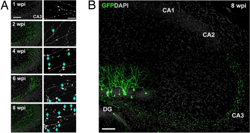
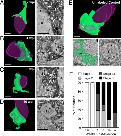
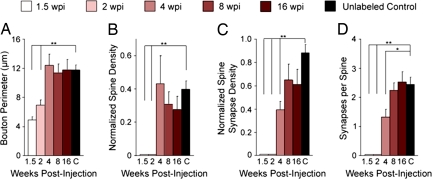
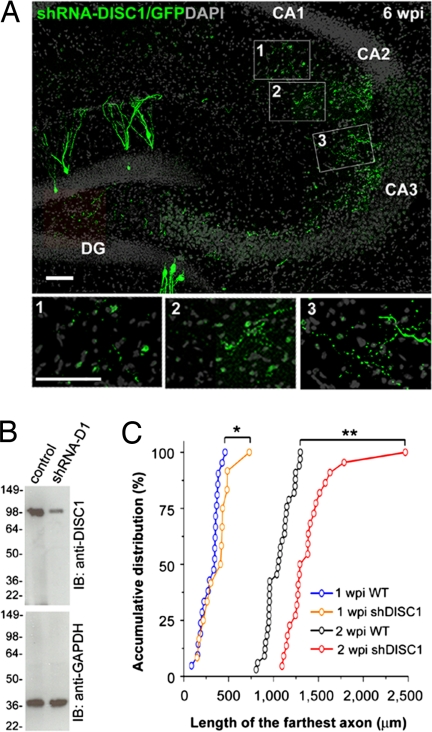
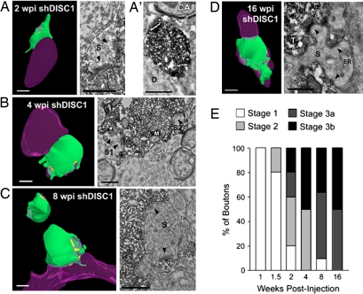
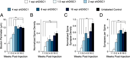
New neurons are continuously generated in restricted regions of the adult mammalian brain. Although these adult-born neurons have been shown to receive synaptic inputs, little is known about their synaptic outputs. Using retrovirus-mediated birth-dating and labeling in combination with serial section electron microscopic reconstruction, we report that mossy fiber en passant boutons of adult-born dentate granule cells form initial synaptic contacts with CA3 pyramidal cells within 2 weeks after their birth and reach morphologic maturity within 8 weeks in the adult hippocampus. Knockdown of Disrupted-in-Schizophrenia-1 (DISC1) in newborn granule cells leads to defects in axonal targeting and development of synaptic outputs in the adult brain. Together with previous reports of synaptic inputs, these results demonstrate that adult-born neurons are fully integrated into the existing neuronal circuitry. Our results also indicate a role for DISC1 in presynaptic development and may have implications for the etiology of schizophrenia and related mental disorders.


Conflict of interest statement

The authors declare no conflict of interest.

Figures

Fig. 1.
Fig. 1.
Axonal development of newborn granule cells in the adult hippocampus. (A) Sample confocal projection images of axons from newborn DGCs in the CA3 subfield at different time points after retroviral labeling. Shown on the left is a low-magnification view of mossy fiber axons (green) and DAPI staining (gray). (Scale bar, 100 μm.) Shown on the right is an enlarged view of axons with expansions (arrows). (Scale bar, 20 μm.) (B) Sample confocal projection image of the hippocampus at 8 wpi. Note that GFP+ mossy fiber axons did not extend beyond the CA3 subfield. (Scale bar, 100 μm.)
Fig. 2.
Fig. 2.
Ultrastructural analysis of adult-born mossy fiber synaptic outputs in the CA3 subfield. (A–D) 3D reconstructions (Left) of serial sections from mossy fiber boutons in the adult mouse with accompanying high-magnification electron micrographs (Right) at 2 wpi (A), 4 wpi (B), 8 wpi (C), and 16 wpi (D). At 2 wpi, synaptic contacts are visible only onto the dendritic shaft (A; arrowheads). Over time, boutons became more mature with synaptic contacts onto dendritic spines (B–D; arrowheads). (E) 3D reconstruction of a mature, unlabeled mossy fiber bouton in the CA3 subfield and accompanying low-magnification (Lower Left) and high-magnification (Lower Right) electron micrographs. The bouton has been pseudocolored in green in the low-magnification image, and the high-magnification image shows synaptic contacts onto an invading dendritic spine (arrowheads). (Scale bars in 3D reconstructions and low-magnification micrograph, 1 μm; in high-magnification micrographs, 0.5 μm.) Green, axonal bouton; purple, dendrite; yellow, synapse. D, dendrite; M, mitochondria; S, dendritic spine; T, axon terminal. (F) Mossy fiber bouton staging paradigm to describe the overall maturation state of boutons across development (see Methods). Shown is the percentage of boutons categorized in each stage for all developmental time points. This suggests that boutons born in the adult brain are qualitatively similar to mature, unlabeled boutons by 8 weeks.
Fig. 3.
Fig. 3.
Quantification of morphologic properties of adult-born mossy fiber boutons. Quantification of average bouton perimeter (A), normalized spine density (B), normalized density of synaptic contacts per bouton onto dendritic spines (C), and number of synaptic contacts per spine (D) at 1.5, 2, 4, 8, and 16 wpi and of unlabeled control boutons (C). This demonstrates that bouton perimeter and spine density are the same as controls by 4 wpi, whereas spinous synaptic contact density and the number of synaptic contacts per spine are the same as controls by 8 wpi. Thus, it takes ≈8 weeks for adult-born mossy fiber boutons to reach morphologic maturity. Values represent mean ± SEM. **P < 0.01; *P < 0.05. See Methods for normalization procedure and refer to Table S1 for raw numbers.
Fig. 4.
Fig. 4.
Axonal development of newborn granule cells with DISC1 knockdown in the adult hippocampus. (A) A sample confocal projection image of mossy fiber axons (green) of newborn neurons with DISC1 knockdown at 6 wpi and DAPI staining (gray). Note the presence of GFP+ axons in the CA1 subfield. (Scale bars, 50 μm.) (B) Knockdown of endogenous full-length DISC1 by shRNA-D1. Primary hippocampal neurons were infected with lentivirus expressing shRNA-D1 and GFP (shRNA-D1), or GFP alone (control), at 2 days in vitro and analyzed at 9 days in vitro by Western blot (IB) using anti-DISC1 antibodies. The membrane was reblotted for GAPDH as a loading control. (C) Quantitative comparison of the length of the farthest axon from WT and shDISC1 mossy fibers at 1 and 2 wpi. Axons with DISC1 knockdown exhibit accelerated outgrowth compared with WT. **P < 0.01; *P < 0.05, Kolmogorov–Smirnov test.
Fig. 5.
Fig. 5.
DISC1 knockdown accelerates adult-born mossy fiber bouton development. (A–D) 3D reconstructions (Left) of serial sections from mossy fiber boutons with accompanying high-magnification electron micrographs (Right) at 2 wpi (A), 4 wpi (B), 8 wpi (C), and 16 wpi (D). At 2 wpi, synaptic contacts (arrowheads) were found on dendritic spines that invaded the mossy fiber bouton (A). Thus, spine invasion and the formation of synaptic contacts were both accelerated with DISC1 knockdown. A low-magnification electron micrograph (A′) shows that in contrast to 2 wpi boutons in CA3 (A), an aberrant bouton in CA1 at 2 wpi lacks synaptic contacts. Boutons appear more mature at later time points (B–D). (Scale bars in 3D reconstructions and low-magnification micrograph, 1 μm; in high-magnification micrographs, 0.5 μm.) Green, axonal bouton; purple, dendrite; yellow, synapse. D, dendrite; ER, endoplasmic reticulum; M, mitochondria; S, dendritic spine; T, axon terminal. (E) Mossy fiber bouton staging paradigm to describe the overall maturation state of boutons across development (see Methods). Shown is the percentage of boutons categorized in each stage for all developmental time points. This demonstrates that, in comparison with WT boutons, the maturation process is accelerated in boutons with DISC1 knockdown, but fewer boutons reach stage 3b at later stages of development.
Fig. 6.
Fig. 6.
Quantification of morphologic properties of adult-born mossy fiber boutons with DISC1 knockdown. Quantification of average bouton perimeter (A), normalized spine density (B), normalized density of synaptic contacts per bouton onto dendritic spines (C), and number of synaptic contacts per spine (D) at 1, 1.5, 2, 4, 8, and 16 wpi and of unlabeled control boutons (C). Control data are repeated from Fig. 3. This demonstrates that bouton perimeter is the same as control by 4 wpi, but the invasion of dendritic spines is accelerated and spine density reaches maturity by 2 wpi. Furthermore, spinous synaptic contact density and the number of synaptic contacts per spine are the same as control by 4 wpi, demonstrating that they reach morphologic maturity faster than WT. Note that boutons at 8 wpi have a significantly smaller spine synapse density than control boutons. Values represent mean ± SEM. **P < 0.01; *P < 0.05. See Methods for normalization procedure and refer to Table S1 for raw numbers.

References

    1. Ming GL, Song H. Adult neurogenesis in the mammalian central nervous system. Annu Rev Neurosci. 2005;28:223–250. - PubMed
    1. Lledo PM, Alonso M, Grubb MS. Adult neurogenesis and functional plasticity in neuronal circuits. Nat Rev Neurosci. 2006;7:179–193. - PubMed
    1. Kitabatake Y, Sailor KA, Ming GL, Song H. Adult neurogenesis and hippocampal memory function: New cells, more plasticity, new memories? Neurosurg Clin N Am. 2007;18:105–113. x. - PMC - PubMed
    1. Zhao C, Deng W, Gage FH. Mechanisms and functional implications of adult neurogenesis. Cell. 2008;132:645–660. - PubMed
    1. van Praag H, et al. Functional neurogenesis in the adult hippocampus. Nature. 2002;415:1030–1034. - PMC - PubMed

Publication types