. 2008 Dec 15;112(13):4884-94.
doi: 10.1182/blood-2008-03-145722. Epub 2008 Sep 9.

Cross-talk of GATA-1 and P-TEFb in megakaryocyte differentiation

Affiliations

Cross-talk of GATA-1 and P-TEFb in megakaryocyte differentiation

Kamaleldin E Elagib et al. Blood. .

Abstract

The transcription factor GATA-1 participates in programming the differentiation of multiple hematopoietic lineages. In megakaryopoiesis, loss of GATA-1 function produces complex developmental abnormalities and underlies the pathogenesis of megakaryocytic leukemia in Down syndrome. Its distinct functions in megakaryocyte and erythroid maturation remain incompletely understood. In this study, we identified functional and physical interaction of GATA-1 with components of the positive transcriptional elongation factor P-TEFb, a complex containing cyclin T1 and the cyclin-dependent kinase 9 (Cdk9). Megakaryocytic induction was associated with dynamic changes in endogenous P-TEFb composition, including recruitment of GATA-1 and dissociation of HEXIM1, a Cdk9 inhibitor. shRNA knockdowns and pharmacologic inhibition both confirmed contribution of Cdk9 activity to megakaryocytic differentiation. In mice with megakaryocytic GATA-1 deficiency, Cdk9 inhibition produced a fulminant but reversible megakaryoblastic disorder reminiscent of the transient myeloproliferative disorder of Down syndrome. P-TEFb has previously been implicated in promoting elongation of paused RNA polymerase II and in programming hypertrophic differentiation of cardiomyocytes. Our results offer evidence for P-TEFb cross-talk with GATA-1 in megakaryocytic differentiation, a program with parallels to cardiomyocyte hypertrophy.


Figures
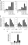
Figure 1
Figure 1
Involvement of P-TEFb in GATA-1 cooperation with RUNX1/CBFβ. (A,B) K562 cells underwent transient transfection with the αIIb-598–luciferase reporter plus expression constructs for GATA-1 (G1), RUNX1 (R1), and CBFβ (Cb) as indicated. Results are mean of 3 independent experiments plus or minus SEM and are expressed as fold activation relative to GATA-1 alone or to vector. All transfections were normalized with cotransfected pCMVβGAL. (A) Transfections also included expression constructs for the dominant-negative mutants Cdk7 KK41/42NQ (dnCdk7) and Cdk9 D167N (dnCdk9). (B) Transfectants were treated the final 24 hours (of 48-hour incubation) with 100, 200, and 300 nM of flavopiridol (FP) or with 25, 50, and 100 μM of DRB. (C) Luciferase reporter assays were conducted as in panel A. K562 transfections included an expression construct for HEXIM1 (HEX) as indicated.
Figure 2
Figure 2
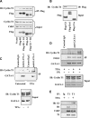
Interaction of GATA-1 with cyclin T1. (A) 293T cells were transfected with constructs encoding either FLAG-RUNX1 (Flag-R1) plus or minus untagged GATA-1 (G1) or FLAG-GATA-1 (Flag-G1) plus or minus untagged RUNX1 (R1). FLAG-immunoprecipitation (IP) was followed by immunoblotting (IB) of immunoprecipitates and inputs with the indicated antibodies. (B) Specificity of interaction. 293T cells transfected with expression vectors for FLAG tagged GATA-1 (G1), Ets1, or C/EBPα (C/α) were subjected to immunoprecipitation and immunoblotting as in panel A. (C) Inducible interaction of endogenous GATA-1 and cycin T1 proteins. Extracts from K562 cells either untreated or treated 48 hours with 25 nM of TPA underwent immunoprecipitation with equal amounts of control rat IgG or N6 monoclonal rat anti-GATA-1 antibody. Immunoprecipitates and inputs underwent immunoblotting with the indicated antibodies. (D) Requirement of P-TEFb kinase activity for inducible recruitment of GATA-1. Coimmunoprecipitations were conducted as in panel C on cells treated 48 hours with 25 nM of TPA plus or minus 100 nM of flavopiridol (FP). (E) Global remodeling of P-TEFb during megakaryocytic induction. Coimmunoprecipitations were conducted as in panel C on cells treated 48 hours with 25 nM of TPA. Immunoprecipitating antibodies consisted of control rabbit IgG or rabbit anticyclin T1 (T1). Immunoblotting was performed for GATA-1 (G1), HEXIM1 (H1), Cdk9, and cyclin T1.
Figure 3
Figure 3
Cdk9 signaling promotes megakaryocytic and inhibits erythroid induction. (A) K562 cells transduced with lentiviral shRNA constructs targeting Cdk9 (C9) underwent immunoblotting of whole cell lysates with the indicated antibodies. (B) Cells from panel A were treated 72 hours with 25 nM of TPA before immunoblotting. (C,D) K562 cells transduced with lentiviral shRNA constructs targeting HEXIM1 (H1) were subjected to immunoblot analysis of the whole cell lysates with the indicated antibodies. R1 indicates RUNX1. Where designated, cells received 200 nM of flavopiridol (FP) for 24 hours before harvesting. (E) Cells from panel C treated with 25 nM of TPA underwent immunoblot analysis. Arrows indicate position of αIIb integrin.
Figure 4
Figure 4
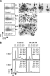
Cdk9 inhibitor interferes with primary human megakaryocyte development. (A,B) Purified human CD34+ cells underwent culture 5 days in unilineage megakaryocytic medium containing TPO, SCF, stromal-derived factor-α, and the indicated doses of flavopiridol (FP). (A) Flow cytometry (FACS) assessment of CD41 expression and forward light scatter (FSC), a reflection of cell size. Analyses were performed on gated viable populations, with percentages of CD41bright FSChi (mature megakaryocytes) and CD41bright FSClo (promegakaryocytes) cells determined using FlowJo software. Wright-stained cytospins were photographed (original magnification ×100). (Top row) Two fields with typical polyploid control megakaryocytes (formula image). (B) Ploidy analysis of CD41+ and CD41 cells within the cultures. Cells were stained with FITC-anti-CD41 and propidium iodide, followed by FACS analysis and quantitation with FlowJo software.
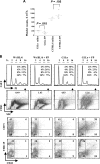
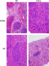
Figure 5
Figure 5
Synthetic interaction of megakaryocyte GATA-1 deficiency with P-TEFb inhibition. (A) GATA-1Lo mice (G1Lo) and age-matched wild-type C57BL/6 received 5 mg/kg per day of flavopiridol (FP; 5 mice per group) or saline control (4 mice per group) for 7 consecutive days. Blood samples from the retro-orbital plexus were then analyzed on a Hemavet automated analyzer for platelet counts. (B) Mice, as in panel A, received a 9-day course of flavopiridol (FP) at the same dose or saline. Marrows were then analyzed by FACS. In rows 1 and 2, marrow cells were costained with anti-CD41 and propidium iodide (PI). The CD41+ cells (percentages indicated in row 2) were gated for ploidy analysis using FlowJo software. Numbers in row 1 indicate percentage of total cells within each ploidy category. In rows 3 and 4, marrow cells were costained antibodies to CD41, TER119 (erythroid marker), and CD71 (transferrin receptor), with quadrant percentages as indicated.
Figure 6
Figure 6
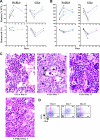
Splenic disruption by megakaryoblastic proliferative disorder. After treatment for 9 days with 5 mg/kg per day of intraperitoneal flavopiridol, mice were killed for histologic analysis of spleens. Comparisons include C57BL/6 (Wt) and GATA-1Lo (G1Lo) strains treated either with saline or flavopiridol (FP). Shown are representative hematoxylin and eosin–stained splenic sections (original magnification ×100).
Figure 7
Figure 7
Reversibility of the megakaryoblastic proliferative disorder. (A-D) C57BL/6 and GATA-1Lo (G1Lo) mice received daily intraperitoneal injection of flavopiridol (7 mg/kg per day) for 7 consecutive days, followed by 2 weeks of no treatment. (A,B) Platelet counts and hematocrits were obtained on days 0, 7, and 21 as indicated. Each line represents an individual mouse. (A,B) Two independent experimental trials. (C) Representative mice were killed at days 0, 7, and 21. Sections from femurs were stained with hematoxylin and eosin (original magnification ×400). Arrows show examples of cells with blastic morphology. (D) Flow cytometry on marrows from mice in panel C.

Comment in

  • GATA-1: one TEF customer.
    Brandt SJ. Brandt SJ. Blood. 2008 Dec 15;112(13):4786-7. doi: 10.1182/blood-2008-10-181743. Blood. 2008. PMID: 19064734 No abstract available.

References

    1. Goldfarb AN. Transcriptional control of megakaryocyte development. Oncogene. 2007;26:6795–6802. - PubMed
    1. Frontelo P, Manwani D, Galdass M, et al. Novel role for EKLF in megakaryocyte lineage commitment. Blood. 2007;110:3871–3880. - PMC - PubMed
    1. Pang L, Weiss MJ, Poncz M. Megakaryocyte biology and related disorders. J Clin Invest. 2005;115:3332–3338. - PMC - PubMed
    1. Shivdasani RA, Fujiwara Y, McDevitt MA, Orkin SH. A lineage-selective knockout establishes the critical role of transcription factor GATA-1 in megakaryocytic growth and platelet development. EMBO J. 1997;16:3965–3973. - PMC - PubMed
    1. Vyas P, Ault K, Jackson CW, Orkin SH, Shivdasani RA. Consequences of GATA-1 deficiency in megakaryocytes and platelets. Blood. 1999;93:2867–2875. - PubMed

Publication types

Substances