

hem6: an ENU-induced recessive hypochromic microcytic anemia mutation in the mouse
- PMID: 18780836
- PMCID: PMC2581980
- DOI: 10.1182/blood-2007-09-111500
hem6: an ENU-induced recessive hypochromic microcytic anemia mutation in the mouse
Abstract
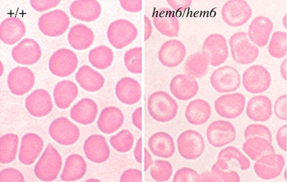
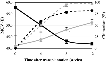
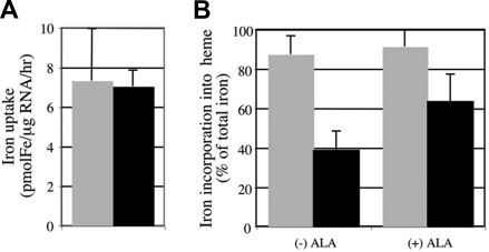
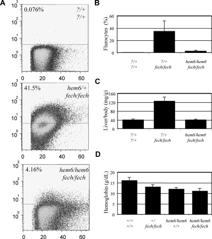
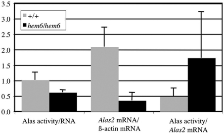
Mouse models have proven invaluable for understanding erythropoiesis. Here, we describe an autosomal recessive, inherited anemia in the mouse mutant hem6. Hematologic and transplantation analyses reveal a mild, congenital, hypochromic, microcytic anemia intrinsic to the hematopoietic system that is associated with a decreased red blood cell zinc protoporphyrin to heme ratio, indicative of porphyrin insufficiency. Intercross matings show that hem6 can suppress the porphyric phenotype of mice with erythropoietic protoporphyria (EPP). Furthermore, iron uptake studies in hem6 reticulocytes demonstrate defective incorporation of iron into heme that can be partially corrected by the addition of porphyrin precursors. Gene expression and enzymatic assays indicate that erythroid 5-aminolevulinic acid synthase (Alas2) is decreased in hem6 animals, suggesting a mechanism that could account for the anemia. Overall, these data lead to the hypothesis that hem6 encodes a protein that directly or indirectly regulates the expression of Alas2.
Figures
Similar articles
-
In ferrochelatase-deficient protoporphyria patients, ALAS2 expression is enhanced and erythrocytic protoporphyrin concentration correlates with iron availability.Blood Cells Mol Dis. 2015 Jan;54(1):71-7. doi: 10.1016/j.bcmd.2014.07.017. Epub 2014 Aug 30. Blood Cells Mol Dis. 2015. PMID: 25179834 Clinical Trial.
-
nm1054: a spontaneous, recessive, hypochromic, microcytic anemia mutation in the mouse.Blood. 2005 Nov 15;106(10):3625-31. doi: 10.1182/blood-2005-01-0379. Epub 2005 Jun 30. Blood. 2005. PMID: 15994289 Free PMC article.
-
Delta-aminolevulinic acid synthase 2 expression in combination with iron as modifiers of disease severity in erythropoietic protoporphyria.Mol Genet Metab. 2019 Nov;128(3):304-308. doi: 10.1016/j.ymgme.2019.04.013. Epub 2019 May 2. Mol Genet Metab. 2019. PMID: 31076252
-
Molecular basis of inherited microcytic anemia due to defects in iron acquisition or heme synthesis.Haematologica. 2009 Mar;94(3):395-408. doi: 10.3324/haematol.13619. Epub 2009 Jan 30. Haematologica. 2009. PMID: 19181781 Free PMC article. Review.
-
[Inheritance in erythropoietic protoporphyria].Pathol Biol (Paris). 2010 Oct;58(5):372-80. doi: 10.1016/j.patbio.2010.01.007. Epub 2010 Sep 20. Pathol Biol (Paris). 2010. PMID: 20850938 Review. French.
Cited by
-
Identification and characterization of a novel murine allele of Tmprss6.Haematologica. 2013 Jun;98(6):854-61. doi: 10.3324/haematol.2012.074617. Epub 2013 Jan 8. Haematologica. 2013. PMID: 23300183 Free PMC article.
-
UBE2O remodels the proteome during terminal erythroid differentiation.Science. 2017 Aug 4;357(6350):eaan0218. doi: 10.1126/science.aan0218. Science. 2017. PMID: 28774900 Free PMC article.
-
Endogenous siderophore 2,5-dihydroxybenzoic acid deficiency promotes anemia and splenic iron overload in mice.Mol Cell Biol. 2014 Jul;34(13):2533-46. doi: 10.1128/MCB.00231-14. Epub 2014 Apr 28. Mol Cell Biol. 2014. PMID: 24777603 Free PMC article.
-
Generation of N-ethyl-N-nitrosourea-induced mouse mutants with deviations in hematological parameters.Mamm Genome. 2011 Oct;22(9-10):495-505. doi: 10.1007/s00335-011-9328-4. Epub 2011 May 8. Mamm Genome. 2011. PMID: 21553221
References
-
- Ney PA. Gene expression during terminal erythroid differentiation. Curr Opin Hematol. 2006;13:203–208. - PubMed
-
- Ingley E, Tilbrook PA, Klinken SP. New insights into the regulation of erythroid cells. IUBMB Life. 2004;56:177–184. - PubMed
-
- Petrak J, Myslivcova D, Man P, Cmejlova J, Cmejla R, Vyoral D. Proteomic analysis of erythroid differentiation induced by hexamethylene bisacetamide in murine erythroleukemia cells. Exp Hematol. 2007;35:193–202. - PubMed
-
- Andrews NC. Animal models of hereditary iron transport disorders. Adv Exp Med Biol. 2002;509:1–17. - PubMed
-
- Ponka P. Tissue-specific regulation of iron metabolism and heme synthesis: distinct control mechanisms in erythroid cells. Blood. 1997;89:1–25. - PubMed
Publication types
MeSH terms
Substances
Grants and funding
LinkOut - more resources
Full Text Sources
Molecular Biology Databases
