. 2008 Sep 9;1(36):ra3.
doi: 10.1126/scisignal.1161577.

Phosphoinositide 3-kinase p110beta activity: key role in metabolism and mammary gland cancer but not development

Affiliations

Phosphoinositide 3-kinase p110beta activity: key role in metabolism and mammary gland cancer but not development

Elisa Ciraolo et al. Sci Signal. .

Abstract

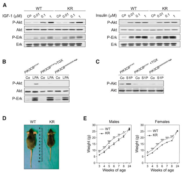
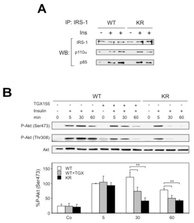
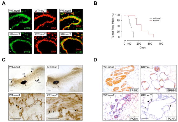
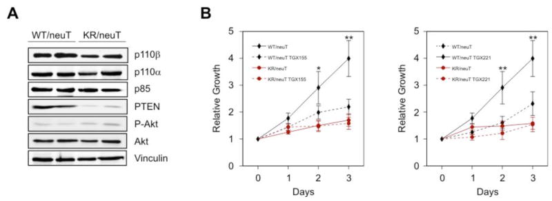
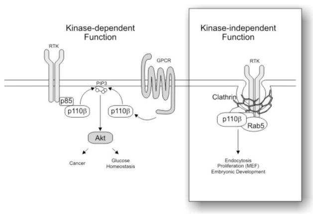
The phosphoinositide 3-kinase (PI3K) pathway crucially controls metabolism and cell growth. Although different PI3K catalytic subunits are known to play distinct roles, the specific in vivo function of p110beta (the product of the PIK3CB gene) is not clear. Here, we show that mouse mutants expressing a catalytically inactive PIK3CB(K805R) mutant survived to adulthood but showed growth retardation and developed mild insulin resistance with age. Pharmacological and genetic analyses of p110beta function revealed that p110beta catalytic activity is required for PI3K signaling downstream of heterotrimeric guanine nucleotide-binding protein (G protein)-coupled receptors as well as to sustain long-term insulin signaling. In addition, PIK3CB(K805R) mice were protected in a model of ERBB2-driven tumor development. These findings indicate an unexpected role for p110beta catalytic activity in diabetes and cancer, opening potential avenues for therapeutic intervention.


Figures

Fig. 1
Fig. 1
Kinase-dependent and independent functions of p110β in wild-type (PIK3CBWT/WT) and PIK3CBK805R/K805R MEFs. A) PIK3CBK805R gene dosage inversely correlates with phenotype severity. MEFs were derived from apparently normal (PIK3CBK805R/K805R High) or abnormal (PIK3CBK805R/K805R Low) 13.5 days post conception (d.p.c.) PIK3CBK805R/K805R embryos. Homogenates of MEFs of the described phenotypes were analyzed by SDS–PAGE and immunoblotted with the indicated antibodies. B) Analysis of p85 association to p110 in MEFs. Protein extracts were immunoprecipitated with anti-pan p85 antibodies and immunoblotted using the indicated antibodies. C) Analysis of p110β catalytic activity. Representative lipid kinase assay with p110β immunoprecipitated from wild-type or PIK3CBK805R/K805R High MEFs. D) PI3K activity in PIK3CBK805R/K805R High (KR) MEFs relative to that in wild type controls (WT), as measured after pull down with anti-p110β antibodies or with phosphopeptide-bound beads associating with all class IA PI3Ks. Asterisks: P < 0.001. E) Proliferation curve of mutant MEFs with high and low p110βK805R expression levels compared to that of wild-type MEFs with or without 100 nM TGX-221 treatment (TGX). Statistical significance: PIK3CBK805R/K805R Low cells vs. all other conditions (* P < 0.05, ** P < 0.01); other pairs of datasets: not significant.
Fig. 2
Fig. 2
Analysis of EGF receptor endocytosis. A) Immunofluorescence of cells of the given genotype incubated with EGF for 1 hour at 4°C (0 min) and then shifted to 37°C for 15 minutes (15 min). Merged images show EGFR in white and 4′-6-Diamidino-2-phenylindole (DAPI) in blue. Exposure times were identical for the different genotypes but longer at 0 min to better show cell surface EGFR. B) Immunofluorescence of cells of the given genotype with anti-clathrin (top) or anti-EEA1 antibodies. Merged images show clathrin or EEA1 in red and DAPI in blue.
Fig. 3
Fig. 3
In vitro and in vivo consequences of p110β kinase-dead expression. A) IGF-1 and insulin-dependent Akt (also known as Protein kinase B, PKB) and extracellular signal regulated kinase 1 and 2 (Erk1/2) phosphorylation in wild-type (WT) and kinase-dead (KR) MEFs derived from normal embryos. Shown is a representative Western blot of eight independent experiments analyzing the response at 5 minutes after stimulation. B) Analysis of Akt phosphorylation after stimulation with LPA. Wild-type (WT) and PIK3CBK805R/K805R High MEFs were stimulated with 10μM LPA in the absence or presence of 100 nM TGX-221. C) Analysis of Akt phosphorylation 5 minutes after stimulation with S1P of cells of the indicated genotype in the presence or absence of the p110β inhibitor TGX221. Shown is a representative Western blot of three independent experiments. D) Growth analysis of PIK3CBK805R/K805R (KR) mice. The figure shows the external appearance of 6 week-old wild-type (WT) and PIK3CBK805R/K805R (KR) mice. E) Weight gain curve of wild-type (WT) and PIK3CBK805R/K805R (KR) mice from 3 to 24 weeks after birth (males n=13, females n=22; * P < 0.05, ** P < 0.01, ** P < 0.001).
Fig. 4
Fig. 4
Insulin-dependent glucose metabolism in 6-month old wild-type (WT) and PIK3CBK805R/K805R mice (KR). Statistical significance: * P < 0.05, ** P < 0.01, ** P < 0.001. A) Blood glucose in random fed mice (n=9 per genotype). B) Insulin concentrations in the serum of random fed mice (n=9 per genotype). C) Blood glucose in fasted mice (wild type, n=9; KR, n=6). D) Glucose tolerance test (n=5 per genotype). E) Insulin tolerance test (n=7 per genotype). F) Insulin concentration in serum of glucose-treated fasted animals. G) Analysis of pancreatic islet size. Left: histological sections of hematoxylin-eosin (H & E) stained pancreata. Center: quantification of islet area (n=4 per genotype). Right: immunofluorescence with antibodies directed against insulin (Ins; red) and against glucagon (Gcn; green). Nuclei were stained with bisbenzimide (DNA; blue). Bars represent 100 μm. H) Histology of a representative liver section of the indicated mice stained with the periodate-Schiff (PAS) stain, recognizing carbohydrates. Measurement of % of PAS positive pixels: WT: 7.3 ± 0.9; KR: 2.27 ± 0.4. I) Glycogen levels in the liver of randomly fed 24 week-old mice (n=7). J) Pyruvate challenge of wild-type (WT) and PIK3CBK805R/K805R (KR) mice. A bolus of 2 g/kg was administered intraperitoneally in 24 week-old mice fasted for 16 hours and blood glucose was measured at the indicated time points (n=5).
Fig. 5
Fig. 5
Role of p110β in insulin signaling. A) Insulin-induced recruitment to IRS-1 of p85 and p110α, determined by IRS-1 immunoprecipitation followed by immunoblot, 5 min after insulin stimulation of livers. B) Phosphorylation of Akt (on Thr308 and Ser473) determined by immunoblot, in livers of mice of the indicated genotype with and without TGX-155 treatment. Lower panel: quantification of Akt phosphorylation on Ser473 (n=5; ** P < 0.01).
Fig. 6
Fig. 6
p110β is required for ERBB2-driven breast cancer development. A) p110β expression in mammary gland epithelium. Cryostat sections of mammary glands derived from PIK3CBK805R/K805R/neuT (KR/neuT) and PIK3CB+/+/neuT (WT) virgin females were stained with the anti-p110β 5g9 monoclonal antibody directly coupled to Alexa 488 (described in Suppl. Fig. 2d; green) as well as with the nuclear stain propidium iodide (red) and analyzed by confocal microscopy. Bar corresponds to 20 μm. B) Kinetics of tumor appearance in PIK3CBWT/WT/neuT (WT/neuT; black lines; n=10) and PIK3CBK805R/K805R/neuT (KR/neuT; red lines; n=7) compound mutant mice (P=0.01). C) Whole mount preparations of PIK3CBWT/WT/neuT and PIK3CBK805R/K805R/neuT mammary glands at 10 weeks. Upper panel: PIK3CBK805R/K805R/neuT mammary gland shows a reduction of duct side buds constituted by atypical hyperplastic lesions. Lower panel: magnification of the side buds revealing the empty aspect of PIK3CBK805R/K805R/neuT hyperplastic lesions. L: lymph node, mh: atypical mammary hyperplasia. Bar corresponds to 1 mm. D) Histology of mammary glands. Ducts were stained with anti Erbb2 and with anti PCNA antibodies to show transgene expression and proliferating cells, respectively. Bar represents 100 μm. Arrowheads indicate PCNA-positive cells in the mutant sample.
Fig. 7
Fig. 7
p110β kinase activity is required for ERBB2-driven breast cancer cell proliferation. A) Western blot analysis of protein expression in cultured mammary tumors of the two genotypes with the indicated antibodies. B) Proliferation curves, reported as relative increase in the number of cells compared to the initial seeding density, of cultured tumor cells of the two genotypes in the absence or presence of the p110β selective inhibitors TGX-155 (10 μM) and TGX-221 (100 nM). Statistical significance: wild-type cells vs. all other conditions (* P < 0.05, ** P < 0.01); other pairs of datasets: not significant.
Fig. 8
Fig. 8
Model of the catalytic and non-catalytic functions of p110β. The catalytic activity of p110β is triggered by both RTK and GPCR signaling and cooperates in Akt activation, cancer development and glucose homeostasis (left side). p110β also functions as a scaffold protein (right inset) required for the organization at the plasma membrane of clathrin coated pits or vesicles, thus controlling RTK endocytosis.

References

    1. Wymann MP, Marone R. Phosphoinositide 3-kinase in disease: timing, location, and scaffolding. Curr Opin Cell Biol. 2005;17:141–149. - PubMed
    1. Engelman JA, Luo J, Cantley LC. The evolution of phosphatidylinositol 3-kinases as regulators of growth and metabolism. Nat Rev Genet. 2006;7:606–619. - PubMed
    1. Hirsch E, Costa C, Ciraolo E. Phosphoinositide 3-kinases as a common platform for multi-hormone signaling. J Endocrinol. 2007;194:243–256. - PubMed
    1. Foukas LC, Claret M, Pearce W, Okkenhaug K, Meek S, Peskett E, Sancho S, Smith AJ, Withers DJ, Vanhaesebroeck B. Critical role for the p110alpha phosphoinositide-3-OH kinase in growth and metabolic regulation. Nature. 2006;441:366–370. - PubMed
    1. Knight ZA, Gonzalez B, Feldman ME, Zunder ER, Goldenberg DD, Williams O, Loewith R, Stokoe D, Balla A, Toth B, Balla T, Weiss WA, Williams RL, Shokat KM. A pharmacological map of the PI3-K family defines a role for p110alpha in insulin signaling. Cell. 2006;125:733–747. - PMC - PubMed

MeSH terms

Substances