Smad7 induces hepatic metastasis in colorectal cancer
- PMID: 18781153
- PMCID: PMC2538763
- DOI: 10.1038/sj.bjc.6604562
Smad7 induces hepatic metastasis in colorectal cancer
Abstract
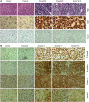
Although Smad signalling is known to play a tumour suppressor role, it has been shown to play a prometastatic function also in breast cancer and melanoma metastasis to bone. In contrast, mutation or reduced level of Smad4 in colorectal cancer is directly correlated to poor survival and increased metastasis. However, the functional role of Smad signalling in metastasis of colorectal cancer has not been elucidated. We previously reported that overexpression of Smad7 in colon adenocarcinoma (FET) cells induces tumorigenicity by blocking TGF-beta-induced growth inhibition and apoptosis. Here, we have observed that abrogation of Smad signalling by Smad7 induces liver metastasis in a splenic injection model. Polymerase chain reaction with genomic DNA from liver metastases indicates that cells expressing Smad7 migrated to the liver. Increased expression of TGF-beta type II receptor in liver metastases is associated with phosphorylation and nuclear accumulation of Smad2. Immunohistochemical analyses have suggested poorly differentiated spindle cell morphology and higher cell proliferation in Smad7-induced liver metastases. Interestingly, we have observed increased expression and junctional staining of Claudin-1, Claudin-4 and E-cadherin in liver metastases. Therefore, this report demonstrates, for the first time, that blockade of TGF-beta/Smad pathway in colon cancer cells induces metastasis, thus supporting an important role of Smad signalling in inhibiting colon cancer metastasis.
Figures


Similar articles
-
Prognostic significance of transforming growth factor beta (TGF-β) signaling axis molecules and E-cadherin in colorectal cancer.Tumour Biol. 2012 Aug;33(4):1005-14. doi: 10.1007/s13277-012-0333-3. Epub 2012 Jan 26. Tumour Biol. 2012. PMID: 22278155
-
PDGF receptor-α promotes TGF-β signaling in hepatic stellate cells via transcriptional and posttranscriptional regulation of TGF-β receptors.Am J Physiol Gastrointest Liver Physiol. 2014 Oct 1;307(7):G749-59. doi: 10.1152/ajpgi.00138.2014. Epub 2014 Aug 28. Am J Physiol Gastrointest Liver Physiol. 2014. PMID: 25169976 Free PMC article.
-
Antimetastatic role of Smad4 signaling in colorectal cancer.Gastroenterology. 2010 Mar;138(3):969-80.e1-3. doi: 10.1053/j.gastro.2009.11.004. Epub 2009 Nov 10. Gastroenterology. 2010. PMID: 19909744 Free PMC article.
-
Transforming growth factor-β signalling: role and consequences of Smad linker region phosphorylation.Cell Signal. 2013 Oct;25(10):2017-24. doi: 10.1016/j.cellsig.2013.06.001. Epub 2013 Jun 11. Cell Signal. 2013. PMID: 23770288 Review.
-
Transforming growth factor-β/Smad signalling in diabetic nephropathy.Clin Exp Pharmacol Physiol. 2012 Aug;39(8):731-8. doi: 10.1111/j.1440-1681.2011.05663.x. Clin Exp Pharmacol Physiol. 2012. PMID: 22211842 Review.
Cited by
-
SMAD7: a timer of tumor progression targeting TGF-β signaling.Tumour Biol. 2014 Sep;35(9):8379-85. doi: 10.1007/s13277-014-2203-7. Epub 2014 Jun 17. Tumour Biol. 2014. PMID: 24935472 Review.
-
Role of TGF-Beta and Smad7 in Gut Inflammation, Fibrosis and Cancer.Biomolecules. 2020 Dec 27;11(1):17. doi: 10.3390/biom11010017. Biomolecules. 2020. PMID: 33375423 Free PMC article. Review.
-
Smad7 Controls Immunoregulatory PDL2/1-PD1 Signaling in Intestinal Inflammation and Autoimmunity.Cell Rep. 2019 Sep 24;28(13):3353-3366.e5. doi: 10.1016/j.celrep.2019.07.065. Cell Rep. 2019. PMID: 31553906 Free PMC article.
-
APOBEC3G promotes liver metastasis in an orthotopic mouse model of colorectal cancer and predicts human hepatic metastasis.J Clin Invest. 2011 Nov;121(11):4526-36. doi: 10.1172/JCI45008. Epub 2011 Oct 10. J Clin Invest. 2011. PMID: 21985787 Free PMC article.
-
Epithelial Claudin Proteins and Their Role in Gastrointestinal Diseases.J Pediatr Gastroenterol Nutr. 2019 May;68(5):611-614. doi: 10.1097/MPG.0000000000002301. J Pediatr Gastroenterol Nutr. 2019. PMID: 30724794 Free PMC article.
References
-
- Afrakhte M, Morén A, Jossan S, Itoh S, Sampath K, Westermark B, Heldin CH, Heldin NE, ten Dijke P (1998) Induction of inhibitory Smad6 and Smad7 mRNA by TGF-beta family members. Biochem Biophys Res Commun 249: 505–511 - PubMed
-
- Agarwal R, D’Souza T, Mortin PJ (2005) Claudin 3 and Claudin 4 expression in ovarian epithelial cells enhances invasion and is associated with increased matrix metalloproteinase-2 activity. Cancer Res 65: 7378–7385 - PubMed
-
- Arnold NB, Ketterer K, Kleeff J, Friess H, Buchler MW, Korc M (2004) Thioredoxin is downstream of Smad7 in a pathway that promotes growth and suppresses cisplatin-induced apoptosis in pancreatic cancer. Cancer Res 64: 3599–3606 - PubMed
-
- Azuma H, Ehata S, Miyazaki H, Watabe T, Maruyama O, Imamura T, Sakamoto T, Kiyama S, Kiyama Y, Ubai T, Inamoto T, Takahara S, Itoh Y, Otsuki Y, Katsuoka Y, Miyazono K, Horie S (2005) Effect of Smad7 expression on metastasis of mouse mammary carcinoma JygMC(A) cells. J Natl Cancer Inst 97: 1734–1746 - PubMed
-
- Behrens J (1999) Cadherins and catenins: role in signal transduction and tumor progression. Cancer Metastasis Rev 18: 15–30 - PubMed
Publication types
MeSH terms
Substances
Grants and funding
LinkOut - more resources
Full Text Sources
Other Literature Sources
Medical
Miscellaneous

