

CD133 expression is an independent prognostic marker for low survival in colorectal cancer
- PMID: 18781171
- PMCID: PMC2570510
- DOI: 10.1038/sj.bjc.6604664
CD133 expression is an independent prognostic marker for low survival in colorectal cancer
Abstract
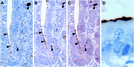
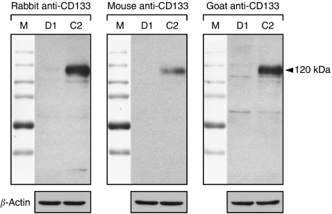
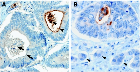
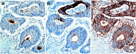
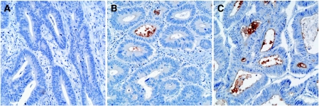
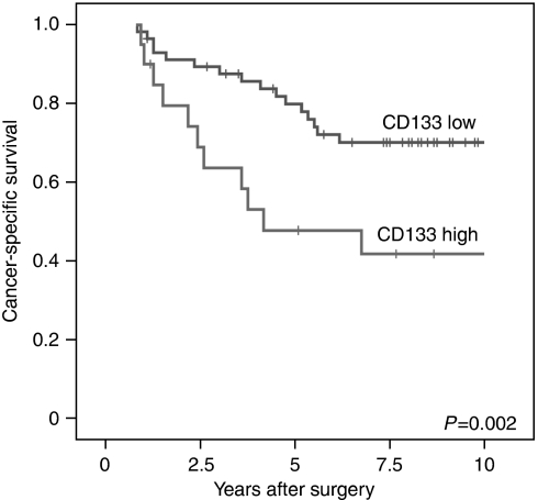
Colon cancer cells have previously been demonstrated to contain a subpopulation of CD133+ tumour cells that have the ability to initiate tumour growth and are thus referred to as colon cancer-initiating cells or colon cancer stem cells (CSCs). As CD133 is currently one of the best markers to characterise colon CSCs, we analysed CD133+ tumour cells in colorectal cancer specimens using immunohistochemistry. We show that CD133 detection is specific and that the CD133 antigen is localised on the glandular-luminal surface of colon cancer cells, whereas undifferentiated tumour cells at the front of invasion are CD133-. In addition, CD133+ cells are characterised in situ by lack of CK20 expression, whereas they are positive for EpCAM. Moreover, we show that CD133 expression in colorectal cancer is an independent prognostic marker that correlates with low survival in a stratified patient collective. Our results indicate that in colorectal cancer, the CD133+ tumour cells can be detected by immunohistochemistry, which facilitates their further characterisation in situ.
Figures
Similar articles
-
Clinicopathological significance and prognostic value of EphA3 and CD133 expression in colorectal carcinoma.J Clin Pathol. 2011 Jun;64(6):498-503. doi: 10.1136/jcp.2010.087213. Epub 2011 Mar 17. J Clin Pathol. 2011. PMID: 21415057
-
Clinical significance of stem cell marker CD133 expression in colorectal cancer.Histol Histopathol. 2016 Mar;31(3):299-306. doi: 10.14670/HH-11-676. Epub 2015 Oct 7. Histol Histopathol. 2016. PMID: 26442717
-
Frequency and pattern of expression of the stem cell marker CD133 have strong prognostic effect on the surgical outcome of colorectal cancer patients.Oncol Rep. 2010 Nov;24(5):1201-12. doi: 10.3892/or_00000973. Oncol Rep. 2010. PMID: 20878111
-
CD133: a cancer stem cells marker, is used in colorectal cancers.World J Gastroenterol. 2013 May 7;19(17):2603-11. doi: 10.3748/wjg.v19.i17.2603. World J Gastroenterol. 2013. PMID: 23674867 Free PMC article. Review.
-
Interconnection of CD133 Stem Cell Marker with Autophagy and Apoptosis in Colorectal Cancer.Int J Mol Sci. 2024 Oct 18;25(20):11201. doi: 10.3390/ijms252011201. Int J Mol Sci. 2024. PMID: 39456981 Free PMC article. Review.
Cited by
-
Tetraspecific scFv construct provides NK cell mediated ADCC and self-sustaining stimuli via insertion of IL-15 as a cross-linker.Oncotarget. 2016 Nov 8;7(45):73830-73844. doi: 10.18632/oncotarget.12073. Oncotarget. 2016. PMID: 27650544 Free PMC article.
-
Simulated microgravity increases polyploid giant cancer cells and nuclear localization of YAP.Sci Rep. 2019 Jul 23;9(1):10684. doi: 10.1038/s41598-019-47116-5. Sci Rep. 2019. PMID: 31337825 Free PMC article.
-
Prognostic impact of the expression of putative cancer stem cell markers CD133, CD166, CD44s, EpCAM, and ALDH1 in colorectal cancer.Br J Cancer. 2010 Jul 27;103(3):382-90. doi: 10.1038/sj.bjc.6605762. Epub 2010 Jul 6. Br J Cancer. 2010. PMID: 20606680 Free PMC article.
-
Surface expression marker profile in colon cancer cell lines and sphere-derived cells suggests complexity in CD26+ cancer stem cells subsets.Biol Open. 2019 Jul 19;8(7):bio041673. doi: 10.1242/bio.041673. Biol Open. 2019. PMID: 31285270 Free PMC article.
-
Advances in Translational Nanotechnology: Challenges and Opportunities.Appl Sci (Basel). 2020;10(14):10.3390/app10144881. doi: 10.3390/app10144881. Appl Sci (Basel). 2020. PMID: 38486792 Free PMC article.
References
-
- Brabletz T, Jung A, Spaderna S, Hlubek F, Kirchner T (2005) Opinion: migrating cancer stem cells – an integrated concept of malignant tumour progression. Nat Rev Cancer 5: 744–749 - PubMed
-
- Burkert J, Wright NA, Alison MR (2006) Stem cells and cancer: an intimate relationship. J Pathol 209: 287–297 - PubMed
-
- Corbeil D, Roper K, Hellwig A, Tavian M, Miraglia S, Watt SM, Simmons PJ, Peault B, Buck DW, Huttner WB (2000) The human AC133 hematopoietic stem cell antigen is also expressed in epithelial cells and targeted to plasma membrane protrusions. J Biol Chem 275: 5512–5520 - PubMed
-
- Hase K, Shatney C, Johnson D, Trollope M, Vierra M (1993) Prognostic value of tumor ‘budding’ in patients with colorectal cancer. Dis Colon Rectum 36: 627–635 - PubMed
Publication types
MeSH terms
Substances
LinkOut - more resources
Full Text Sources
Other Literature Sources
Medical
Research Materials
Miscellaneous
