. 2008 Sep;41(6):442-53.
doi: 10.1080/08916930802031579.

Consequences of unlocking the cardiac myosin molecule in human myocarditis and cardiomyopathies

Affiliations

Consequences of unlocking the cardiac myosin molecule in human myocarditis and cardiomyopathies

Adita Mascaro-Blanco et al. Autoimmunity. 2008 Sep.

Abstract

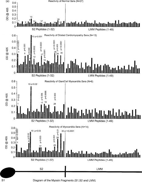
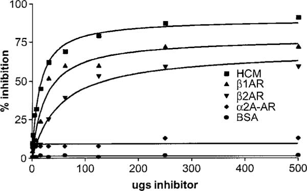
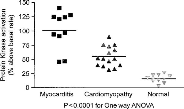
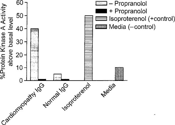
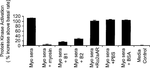
Myocarditis, often initiated by viral infection, may progress to autoimmune inflammatory heart disease, dilated cardiomyopathy and heart failure. Although cardiac myosin is a dominant autoantigen in animal models of myocarditis and is released from the heart during viral myocarditis, the characterization, role and significance of anti-cardiac myosin autoantibodies is poorly defined. In our study, we define the human cardiac myosin epitopes in human myocarditis and cardiomyopathies and establish a mechanism to explain how anti-cardiac myosin autoantibodies may contribute to heart disease. We show that autoantibodies to cardiac myosin in sera from myocarditis and dilated cardiomyopathies in humans targeted primarily epitopes in the S2 hinge region of cardiac myosin. In addition, anti-cardiac myosin antibodies in sera or purified IgG from myocarditis and cardiomyopathy targeted the beta-adrenergic receptor and induced antibody-mediated cAMP-dependent protein kinase A (PKA) cell signaling activity in heart cells. Antibody-mediated PKA activity in sera was abrogated by absorption with anti-human IgG. Antibody-mediated cell signaling of PKA was blocked by antigen-specific inhibition by human cardiac myosin or the beta-adrenergic receptor but not the alpha adrenergic receptor or bovine serum albumin. Propranolol, a beta blocker and inhibitor of the beta-adrenergic receptor pathway also blocked the antibody-mediated signaling of the beta-adrenergic receptor and PKA. The data suggest that IgG antibody against human cardiac myosin reacts with the beta-adrenergic receptor and triggers PKA signaling in heart cells. In summary, we have identified a new class of crossreactive autoantibodies against human cardiac myosin and the beta-adrenergic receptor in the heart. In addition, we have defined disease specific peptide epitopes in the human cardiac myosin rod S2 region in human myocarditis and cardiomyopathy as well as a mechanistic role of autoantibody in the pathogenesis of disease.


Figures

Figure 1
Figure 1
Anti-human cardiac myosin titers (IgG) in sera from myocarditis (p ≤ 0.0001) were significantly higher compared with cardiomyopathy and normal control sera in the ELISA.
Figure 2
Figure 2
Anti-human cardiac myosin titers (IgG) of diabetic cardiomyopathy sera compared with diabetic sera and normal control donors in the ELISA. Diabetic cardiomyopathy sera titers were significantly higher than in diabetic (p ≤ 0.0001) and control (p = 0.01) sera groups.
Figure 3
Figure 3
(A) Comparison of sera from myocarditis, dilated cardiomyopathy, giant cell myocarditis and normal control patients in their reactivity with 82 synthetic peptides spanning the S2 (25-mers) hinge region and LMM (18-mers) tail region of the human cardiac myosin rod in the ELISA (sera @ 1/100 dilution). Significant reactivity (OD) compared to normal controls for the peptides is indicated with p values and grey bars. (B) Comparison diabetic, diabetic cardiomyopathy, and normal sera reactivity with S2 and LMM synthetic peptides. Sera from the control donors rarely reacted with S2 peptides at an OD >0.2. Significant reactivity compared to normal controls is indicated by p values and grey bars.
Figure 3
Figure 3
(A) Comparison of sera from myocarditis, dilated cardiomyopathy, giant cell myocarditis and normal control patients in their reactivity with 82 synthetic peptides spanning the S2 (25-mers) hinge region and LMM (18-mers) tail region of the human cardiac myosin rod in the ELISA (sera @ 1/100 dilution). Significant reactivity (OD) compared to normal controls for the peptides is indicated with p values and grey bars. (B) Comparison diabetic, diabetic cardiomyopathy, and normal sera reactivity with S2 and LMM synthetic peptides. Sera from the control donors rarely reacted with S2 peptides at an OD >0.2. Significant reactivity compared to normal controls is indicated by p values and grey bars.
Figure 4
Figure 4
Dose response curves demonstrate antigen-specific inhibition of IgG reactivity with the beta-1 adrenergic receptor by human cardiac myosin and B1 or B2 adrenergic receptors. Reactivity of purified IgG from cardiomyopathy sera is inhibited by human cardiac myosin, and B1 or B2 adrenergic receptors but not by the alpha 2 adrenergic receptor or BSA.
Figure 5
Figure 5
Antibody-mediated cell signaling by sera from myocarditis and cardiomyopathy. H9c2 heart cells in culture were treated with sera (1:100 dilution) or purified IgG (100 μg/ml) and illustrate the levels of PKA signaling in the heart cells.
Figure 6
Figure 6
Propranolol, a beta-adrenergic receptor antagonist, is shown to effectively inhibit the PKA activity of the purified IgG from selected cardiomyopathy sera. Purified IgG from cardiomyopathy activated PKA significantly above basal PKA levels while purified normal IgG did not. H9c2 heart cells in culture were treated with purified IgG and PKA activity measured in counts per minute as described in materials and methods.
Figure 7
Figure 7
Activation of PKA by IgG in myocarditis serum. Anti-IgG beads remove the antibody-mediated PKA activity while BSA coated beads do not. Therefore, autoantibodies which mediate cell signaling in cardiomyocytes can be blocked by anti-IgG.
Figure 8
Figure 8
Autoantibodies which can mediate PKA cell signaling in cardiomyocytes can be blocked by the specific antigens human cardiac myosin and beta-adrenergic receptors. Antibodies do not react with the Rα2aAR or BSA. Antigen-specific inhibition pattern was similar to cardiomyopathy sera compared to myocarditis sera as shown in PKA assay.

Similar articles

Cited by

References

    1. Caforio ALP, Goldman JH, Haven AJ, Baig KM, McKenna WJ. Evidence for autoimmunity to myosin and other heart-specific autoantigens in patients with dilated cardiomyopathy and their relatives. Int J Cardiol. 1996;54:157–163. - PubMed
    1. Brown CA, O'Connell JB. Myocarditis and idiopathic dilated cardiomyopathy. Am J Med. 1995;99:309–314. - PMC - PubMed
    1. Felker GM, Hu W, Hare JM, Hruban RH, Baughman KI, Kasper EK. The spectrum of dilated cardiomyopathy. The Johns Hopkins experience with 1278 patients. Medicine. 1999;78:270–283. - PubMed
    1. Aretz HT. Myocarditis: The Dallas criteria. Hum Pathol. 1987;18:619–624. - PubMed
    1. Aretz HT, Billingham ME, Edwards WD, Factor SM, Fallon JT, Fenoglio JJ, Jr, et al. Myocarditis. A histopathologic definition and classification. Am J Cardiovasc Pathol. 1987;1:3–14. - PubMed

Publication types

MeSH terms