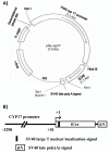
Generation of Cyp17iCre transgenic mice and their application to conditionally delete estrogen receptor alpha (Esr1) from the ovary and testis
- PMID: 18781648
- PMCID: PMC2637183
- DOI: 10.1002/dvg.20428
Generation of Cyp17iCre transgenic mice and their application to conditionally delete estrogen receptor alpha (Esr1) from the ovary and testis
Abstract
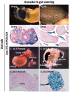
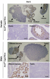
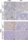
A transgenic mouse line that expresses iCre under regulation of the Cytochrome P(450) 17alpha-hydroxylase/17, 20-lyase (Cyp17) promoter was developed as a novel transgenic mouse model for the conditional deletion of genes specifically in the theca/interstitial cells of the ovary and Leydig cells of the testis. In this report, we describe the development of Cyp17iCre mice and the application of these mice for conditional deletion of the estrogen receptor alpha (Esr1) gene in the theca/interstitial and Leydig cells of the female and male gonad, respectively. These mice will prove a powerful tool to inactivate genes in the gonad in a cell-specific manner.
Figures


Similar articles
-
Inhibin alpha-iCre mice: Cre deleter lines for the gonads, pituitary, and adrenal.Genesis. 2006 Apr;44(4):183-8. doi: 10.1002/dvg.20198. Genesis. 2006. PMID: 16604527
-
Generation of an estrogen receptor beta-iCre knock-in mouse.Genesis. 2016 Jan;54(1):38-52. doi: 10.1002/dvg.22911. Epub 2016 Jan 8. Genesis. 2016. PMID: 26663382 Free PMC article.
-
Generation and characterization of an estrogen receptor alpha-iCre knock-in mouse.Genesis. 2017 Dec;55(12):10.1002/dvg.23084. doi: 10.1002/dvg.23084. Epub 2017 Nov 17. Genesis. 2017. PMID: 29115049 Free PMC article.
-
17α-Hydroxylase (CYP17) expression and subsequent androstenedione production in the human ovary.Reprod Sci. 2010 Nov;17(11):978-86. doi: 10.1177/1933719110379055. Epub 2010 Aug 18. Reprod Sci. 2010. PMID: 20720262 Review.
-
Origin and Differentiation of Androgen-Producing Cells in the Gonads.Results Probl Cell Differ. 2016;58:101-34. doi: 10.1007/978-3-319-31973-5_5. Results Probl Cell Differ. 2016. PMID: 27300177 Review.
Cited by
-
Lrh1 can help reprogram sexual cell fate and is required for Sertoli cell development and spermatogenesis in the mouse testis.PLoS Genet. 2022 Feb 22;18(2):e1010088. doi: 10.1371/journal.pgen.1010088. eCollection 2022 Feb. PLoS Genet. 2022. PMID: 35192609 Free PMC article.
-
Leydig cell genes change their expression and association with polysomes in a stage-specific manner in the adult mouse testis.Biol Reprod. 2018 May 1;98(5):722-738. doi: 10.1093/biolre/ioy031. Biol Reprod. 2018. PMID: 29408990 Free PMC article.
-
Retinoic acid receptor signaling is necessary in steroidogenic cells for normal spermatogenesis and epididymal function.Development. 2018 Jul 9;145(13):dev160465. doi: 10.1242/dev.160465. Development. 2018. PMID: 29899137 Free PMC article.
-
A Novel Model Using AAV9-Cre to Knockout Adult Leydig Cell Gene Expression Reveals a Physiological Role of Glucocorticoid Receptor Signalling in Leydig Cell Function.Int J Mol Sci. 2022 Nov 30;23(23):15015. doi: 10.3390/ijms232315015. Int J Mol Sci. 2022. PMID: 36499341 Free PMC article.
-
Aberrant activation of KRAS in mouse theca-interstitial cells results in female infertility.Front Physiol. 2022 Aug 19;13:991719. doi: 10.3389/fphys.2022.991719. eCollection 2022. Front Physiol. 2022. PMID: 36060690 Free PMC article.
References
-
- Arensburg J, Payne AH, Orly J. Expression of Steroidogenic Genes in Maternal and Extraembryonic Cells During Early Pregnancy in Mice. Endocrinology. 1999;140:5220–5232. - PubMed
-
- Bell P, Limberis M, Gao G, Wu D, Bove MS, Sanmiguel JC, Wilson JM. An optimized protocol for detection of E. coli β-galactosidase in lung tissue following gene transfer. Histochem Cell Biol. 2005;124:77–85. - PubMed
-
- Bingham NC, Verma-Kurvari S, Parada LF, Parker KL. Development of a steroidogenic factor 1/Cre transgenic mouse line. Genesis. 2006;44:419–424. - PubMed
-
- Dupont S, Krust A, Gansmuller A, Dierich A, Chambon P, Mark M. Effect of single and compound knockouts of estrogen receptors a (ERa) and b (ERb) on mouse reproductive phenotypes. Development. 2000;127:4277–4291. - PubMed
-
- Durkee TJ, McLean MP, Hales DB, Payne AH, Waterman MR, Khan I, Gibori G. P450(17 alpha) and P450SCC gene expression and regulation in the rat placenta. Endocrinology. 1992;130:1309–1317. - PubMed
Publication types
MeSH terms
Substances
Grants and funding
LinkOut - more resources
Full Text Sources
Molecular Biology Databases
Research Materials
Miscellaneous

