. 2008 Nov;154(2):162-71.
doi: 10.1111/j.1365-2249.2008.03762.x. Epub 2008 Sep 8.

Effect of proteolytic activity of Epicoccum purpurascens major allergen, Epi p 1 in allergic inflammation

Affiliations

Effect of proteolytic activity of Epicoccum purpurascens major allergen, Epi p 1 in allergic inflammation

N Kukreja et al. Clin Exp Immunol. 2008 Nov.

Abstract

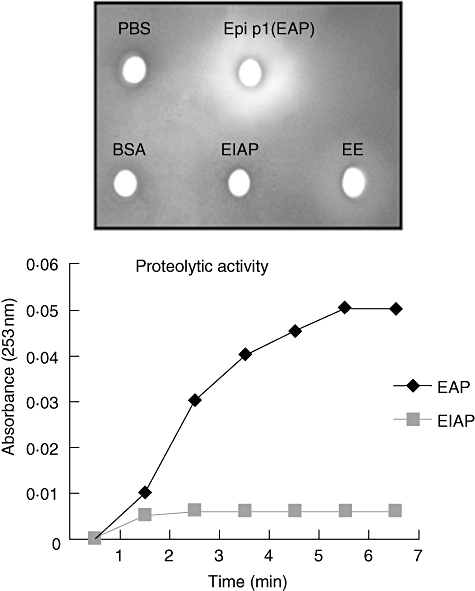
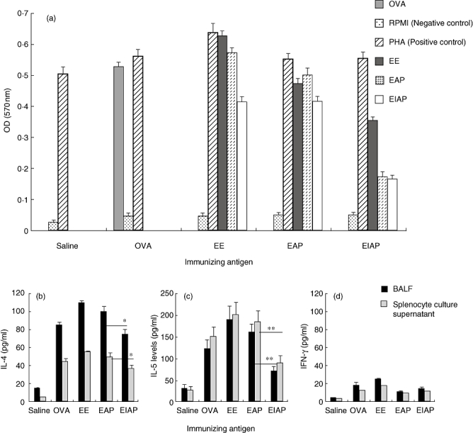
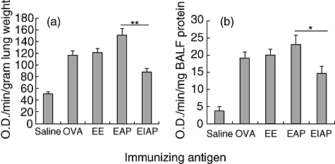
Enzymes play an important role in inducing airway inflammation, but knowledge is limited to few proteins. This study was carried out to assess the role of Epi p 1, a serine protease of Epicoccum purpurascens, in inducing allergy and inflammation in a murine model. Balb/c mice were sensitized with Epi p 1 active protease (EAP) or Epicoccum extract. Subsequently, Epi p 1 sensitized mice were boosted on day 14 with EAP or inactivated protease (EIAP). Three intranasal challenges were given and mice were killed to obtain blood, bronchoalveolar lavage fluid (BALF), spleen and lung tissues. Cellular airways infiltration, immunoglobulin E (Ig)E titres and cytokine levels in BALF and splenocyte culture supernatant were compared. Mice immunized with EAP had higher Epi p 1-specific serum IgE and IgG1 than EIAP immunized mice (P < 0.01). There was a twofold difference in the number of eosinophils in BALF of EAP mice and EIAP mice (P < 0.01). A similar trend was recorded for eosinophil peroxidase activity (P < 0.05), indicating the role of proteolytic activity in inducing inflammation. Further, lung histology revealed increased leucocyte infiltration and airway narrowing, with higher inflammation scores in the EAP group than in the EIAP group. The lungs of EAP mice showed increased mucus and goblet cell metaplasia. Interleukin (IL)-4 and IL-5 levels were higher in BALF and splenocyte culture supernatant of EAP mice than in EIAP mice (P < 0.05), indicating a T helper 2 response. Proteolytic activity of Epi p 1 plays an important role in inducing allergic inflammation. The enzymatically inactive form may be investigated for immunotherapy.


Figures

Fig. 1
Fig. 1
Enzymatic activity of Epi p 1. (a) Hydrolytic activity of Epi p 1 active protease (EAP) or phenyl methyl sulphonyl fluoride (PMSF) treated, i.e. inactivated, protease and Epicoccum extract (EE) was checked by agarose plate assay. Phosphate-buffered saline (PBS) and bovine serum albumin (BSA) were used as negative controls. Each antigen was incubated in wells of 0·1% agarose gel containing 1% gelatin. Proteolytic activity was recorded as clear zone on Coomassie brilliant blue-stained gel. (b) Active protease (EAP) and PMSF-treated, i.e. inactivated protease, were assayed by Nα-benzoyl-L-arginine-ethyl ester hydrochloride hydrolysis as an increase in absorbance per unit time. There was a statistically significant difference in hydrolytic activity of EAP and EIAP.
Fig. 2
Fig. 2
Serum antibody responses to ovalbumin (OVA), Epicoccum extract (EE), Epi p 1 active protease (EAP) and Epi p 1 inactivated protease (EIAP), prior to and after sensitization with respective antigens. The immunoglobulin E (IgE), IgG1 levels and IgG2a were detected using appropriate secondary antibodies in enzyme-linked immunosorbent assay (ELISA). Average absorbance value of six mice in each group is plotted with standard deviation (s.d.). Horizontal line (formula image
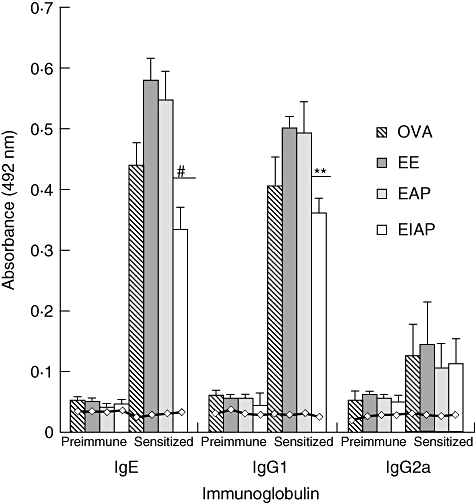
) shows the values of saline control in each group. Statistical significance between specific IgE/IgG1 of EAP and EIAP sensitized mice is mentioned as P-value (at a confidence level of 0·05); #P < 0·005; **P < 0·01. Data are presented as ± s.d.
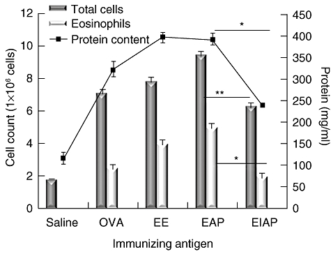
Fig. 3
Fig. 3
Effects of administration of various antigens on total cell and eosinophil number (primary axis) and protein content (secondary axis) in BALF. Saline, ovalbumin (OVA), Epicoccum extract (EE), Epi p 1 active protease (EAP) and Epi p 1 inactivated protease (EIAP) were administered to the mice in different groups. The protein concentration (shown as line plotted) was measured by Bradford assay. The total number of cells was counted using a haemocytometer. The eosinophils were counted on cell smears stained with Leishman's stain. Statistical difference in each parameter of EAP and EIAP sensitized mice is mentioned as P-value (at a confidence level of 0·05); *P < 0·05; **P < 0·01.
Fig. 4
Fig. 4
Effect of various antigens on (a) splenocyte proliferation, (b) interleukin (IL)-4, (c) IL-5 and (d) interferon (IFN)-γ release. Spleen cells of mice from different groups were stimulated with Epicoccum extract (EE), Epi p 1 active protease (EAP) and/or Epi p 1 inactivated protease (EIAP). The cells from the ovalbumin (OVA) immunized group were stimulated with OVA. Phytohaemagglutinin and RPMI-1640 were used as positive and negative controls respectively. The cell proliferation was measured by 3-(4, 5-dimethylthiazol-2-yl)-2, 5-diphenyltetrazolium bromide assay after 72 h of incubation at 37°C in a CO2 incubator. Cytokines were measured by enzyme-linked immunosorbent assay. Statistical significance between cytokine levels of EAP and EIAP sensitized mice is mentioned as P-value (at a confidence level of 0·05); *P < 0·05; **P < 0·01.
Fig. 5
Fig. 5
Light photomicrographs of lungs of mice treated with various antigens (20 × resolution). (a) Haematoxylin and eosin-stained paraffin section of lung tissue; (b) periodic acid-Schiff (PAS)-stained paraffin section of lung tissue. The mice were exposed to saline and antigens, e.g. OVA, Epicoccum extract (EE), Epi p 1 active protease (EAP) and Epi p 1 inactivated protease (EIAP). Mice immunized with OVA, EE and EAP showed increased cellular infiltration and the obstruction of airways with mucus. Inset on the top left of each figure shows area marked by arrow at higher resolution (40×).The infiltration score and goblet cell metaplasia score is shown with each staining. Statistical difference in each parameter of EAP and EIAP sensitized mice is mentioned as P-value (at a confidence level of 0·05); *P < 0·05.
Fig. 6
Fig. 6
Eosinophil peroxidase (EPO) activity in (a) lung homogenate and (b) bronchoalveolar lavage fluid (BALF) of saline, ovalbumin (OVA), Epicoccum extract (EE), Epi p 1 active protease (EAP) and Epi p 1 inactivated protease (EIAP) treated mice. Change in absorbance of substrate in wells containing BALF or lung homogenate from the above-mentioned groups was recorded as EPO activity. Statistical difference in each parameter of EAP and EIAP sensitized mice is mentioned as P-value (at a confidence level of 0·05); *P < 0·05; **P < 0·01.

Similar articles

Cited by

References

    1. Jeong SK, Kim HJ, Youm JK, et al. Mite and cockroach allergens activate protease activated receptor-2 and delay epidermal permeability barrier recovery. J Invest Dermatol. 2008;128:1930–9. - PubMed
    1. Runswick S, Mitchell T, Davies P, Robinson C, Garrod DR. Pollen proteolytic enzymes degrade tight junctions. Respirology. 2007;12:834–42. - PubMed
    1. Shen HD, Tam MF, Tang RB, Chou H. Aspergillus and Penicillium allergens: focus on proteases. Curr Allergy Asthma Rep. 2007;7:351–6. - PubMed
    1. Ghaemmaghami AM, Gough L, Sewell HF, Shakib F. The proteolytic activity of the major dust mite allergen Der p 1 conditions dendritic cells to produce less interleukin-12: allergen-induced Th2 bias determined at the dendritic cell level. Clin Exp Allergy. 2002;32:1468–75. - PubMed
    1. Gough L, Schulz O, Sewell HF, Shakib F. The cysteine protease activity of the major dust mite allergen Der p 1 selectively enhances the immunoglobulin E antibody response. J Exp Med. 1999;190:1897–902. - PMC - PubMed

Publication types

MeSH terms