. 2008 Dec;154(3):365-74.
doi: 10.1111/j.1365-2249.2008.03757.x. Epub 2008 Sep 8.

Generation of functionally mature dendritic cells from elutriated monocytes using polyinosinic : polycytidylic acid and soluble CD40 ligand for clinical application

Affiliations

Generation of functionally mature dendritic cells from elutriated monocytes using polyinosinic : polycytidylic acid and soluble CD40 ligand for clinical application

S Kim et al. Clin Exp Immunol. 2008 Dec.

Abstract

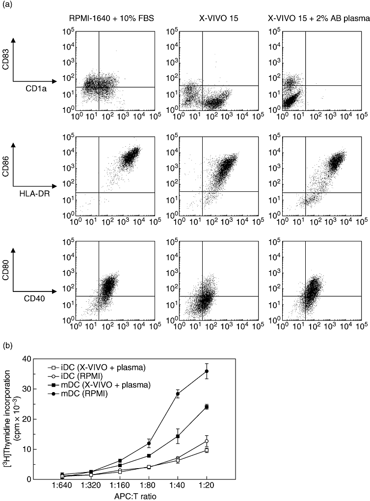
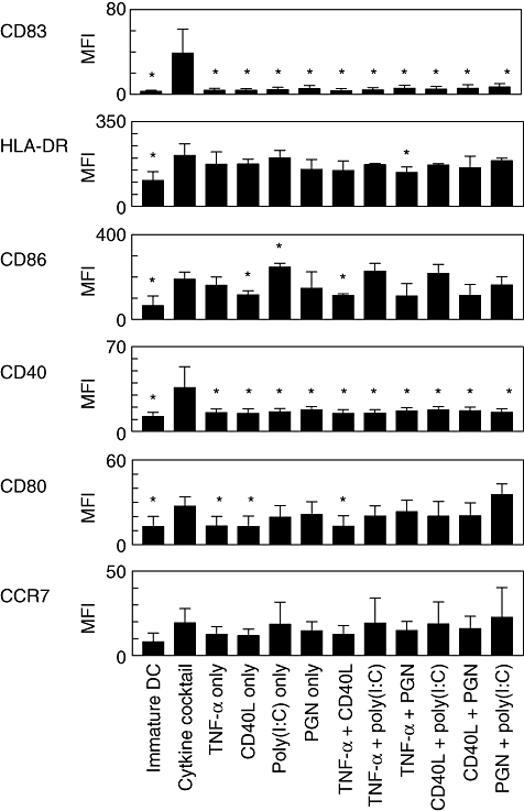
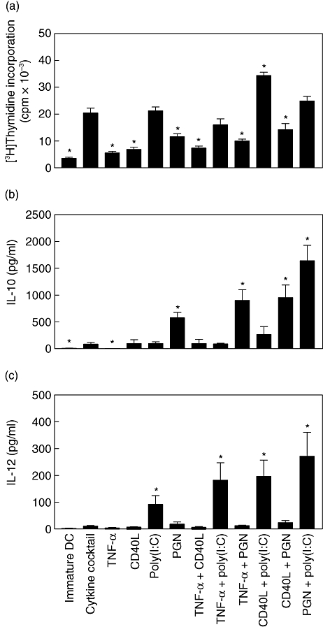
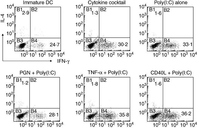
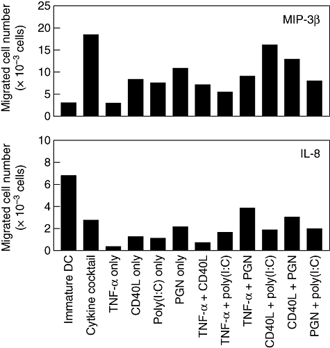
Despite the increasing use of dendritic cell (DC) vaccination in clinical trials, optimal conditions for the generation of functionally mature DCs remain to be established. The current standard DC maturation protocol for clinical trials has been used as an inflammatory cytokine cocktail [tumour necrosis factor (TNF)-alpha, interleukin (IL)-1beta, IL-6 and prostaglandin E(2)], but this cocktail induced insufficient maturation of DCs derived from elutriated monocytes when cultured in X-VIVO 15. The aim of this study was to define effective combinations of stimulators for generating functionally mature DCs from elutriated monocytes under current good manufacturing practice conditions. We compared the functional capacity of DCs in response to all possible pairwise combinations of four different classes of stimuli: TNF-alpha, peptidoglycan, polyinosinic : polycytidylic acid [poly(I:C)] and soluble CD40 ligand (CD40L). Maturation status of DCs stimulated with combination of four stimuli was similar to that of the cytokine cocktail as assessed by the cell surface phenotype. However, only the combination of poly(I:C) + CD40L induced complete functional activation of the whole DC population, assessing IL-12p70 production, allostimulatory activity, migratory response to CCL19 and T helper 1-polarizing capacity. Thus, the protocol based on the combination of poly(I:C) and CD40L is more effective for the induction of clinical-grade DCs from elutriated monocytes than the standard cytokine cocktail.


Figures

Fig. 1
Fig. 1
Phenotype and allostimulatory activity of mature dendritic cells (mDCs) generated from elutriated monocytes. (a) Elutriated monocyte-derived immature DCs (iDCs) were stimulated with tumour necrosis factor (TNF)-α, interleukin (IL)-1β, IL-6 and prostaglandin E2(PGE2) for 48 h in the presence of RPMI-1640 + 10% fetal bovine serum (FBS), serum-free X-VIVO 15 and X-VIVO 15 with 2% human AB plasma. Expression of CD83, CD1a, human leucocyte antigen D-related (HLA-DR), CD86, CD40 and CD80 was analysed by flow cytometry. Compared with the DCs cultured with RPMI-1640 + 10% FBS, DCs cultured with X-VIVO 15 expressed low level of CD83. The representative result of one1 of four experiments is shown. (b) Allogeneic mixed leucocyte reaction assay. The mDCs were generated in the presence of RPMI-1640 + 10% FBS or X-VIVO 15 + 2% human AB plasma stimulated with TNF-α, IL-1β, IL-6 and PGE2. Allogeneic peripheral blood mononuclear cells (105 cells/well) as responders were stimulated with graded numbers of mature and immature DCs (ratio ranging between 1:20 and 1:640). Proliferation was measured by [3H]-thymidine incorporation on day 5. Results from one representative of three independent experiments are shown.
Fig. 2
Fig. 2
Phenotypic profiles of dendritic cells (DCs) stimulated with different combinations of maturation factors. Immature DCs were stimulated for 48 h with single or pairwise combinations of tumour necrosis factor (TNF)-α, CD40L, polyinosinic : polycytidylic acid [poly(I:C)] and peptidoglycan (PGN), and maturation status was assessed by expression of cell surface markers (CD83, HLA-DR, CD86, CD40, CD80 and CCR7). As a positive control, matured DCs matured with cytokine cocktail (TNF-α, interleukin-1β, IL-6 and prostaglandin E2) were included. Values represent mean fluorescence intensity (MFI) ± standard error of the mean from three independent experiments. *Increased or decreased MFI with respect to control (DCs matured with cytokine cocktail) is statistically significant (P < 0·05).
Fig. 3
Fig. 3
Allogeneic T cell proliferation and cytokine secretion by dendritic cells (DCs) matured with different combinations of maturation stimuli. (a) Capacity of mature DCs (mDCs) generated with different combinations of maturation stimuli to induce allogeneic T cell proliferation. Immature DCs were stimulated with the indicated maturation stimuli for 48 h, and the mDCs were used to stimulate proliferation of allogeneic peripheral blood mononuclear cells (2 × 10⁵ cells/well) at different stimulator : responder ratios. Data from the stimulator : responder ratio at 1:10 are presented as mean counts of incorporated [3H]-thymidine. Results shown represent the mean ± standard error of the mean (s.e.m.) of four independent experiments. *Increased or decreased cell proliferation with respect to control (DCs matured with cytokine cocktail) is statistically significant (P < 0·05). (b) IL-10 and (c) IL-12p70 were measured in culture supernatants 48 h later. Results are expressed as mean ± s.e.m. of six independent experiments. *Increased cytokine production with respect to control (DCs matured with cytokine cocktail) is statistically significant (P < 0·05).
Fig. 4
Fig. 4
Intracellular cytokine expression of dendritic cell (DC)-activated T cells. Immature DCs, DCs matured with either cytokine cocktail, polyinosinic : polycytidylic acid [poly(I:C)] (20 µg/ml) alone or a combination of poly(I:C) (20 µg/ml) with tumour necrosis factor-α (10 ng/ml), peptidoglycan (10 µg/ml) and CD40L (1 µg/ml) were co-cultured with allogeneic CD4+ T cells. After 7 days of co-culture, T cells were restimulated with phorbol 12-myristate 13-acetate and ionomycin for 8 h. Brefeldin A was added to the culture during the last 6 h before staining to prevent cytokine release. Quadrants were set according to the fluorescence intensities of isotype-matched control antibodies with irrelevant specificity. Results from one representative of three independent experiments are shown.
Fig. 5
Fig. 5
Chemotactic response of dendritic cells (DCs) in response to major inflammatory protein (MIP)-3β and interleukin (IL)-8. DCs (1 × 10⁵) were seeded in the upper compartment of Transwell plates and incubated for 2 h at 37°C in the presence or absence of MIP-3β (10 ng/ml) or IL-8 (50 ng/ml) added to the lower compartment. DC counts in the lower chamber wells were measured by flow cytometry. Chemotaxis in the absence of any chemokines (medium) was the negative control. Results from one representative of three independent experiments are shown.

Similar articles

Cited by

References

    1. Banchereau J, Steinman RM. Dendritic cells and the control of immunity. Nature. 1998;392:245–52. - PubMed
    1. Osada T, Clay TM, Woo CY, Morse MA, Lyerly HK. Dendritic cell-based immunotherapy. Int Rev Immunol. 2006;25:377–413. - PubMed
    1. Fong L, Engleman EG. Dendritic cells in cancer immunotherapy. Ann Rev Immunol. 2000;18:245–73. - PubMed
    1. Whiteside TL, Odoux C. Dendritic cell biology and cancer therapy. Cancer Immunol Immunother. 2004;53:240–8. - PMC - PubMed
    1. O'Neill DW, Adams S, Bhardwaj N. Manipulating dendritic cell biology for the active immunotherapy of cancer. Blood. 2004;104:2235–46. - PubMed

Publication types