. 2008 Nov 14;283(46):31991-2002.
doi: 10.1074/jbc.M803612200. Epub 2008 Sep 9.

SUMOylation regulates nuclear localization of Krüppel-like factor 5

Affiliations

SUMOylation regulates nuclear localization of Krüppel-like factor 5

James X Du et al. J Biol Chem. .

Abstract

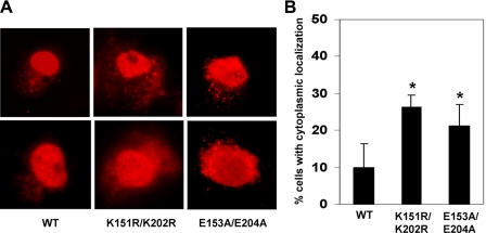
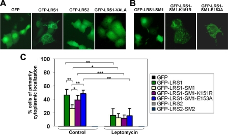
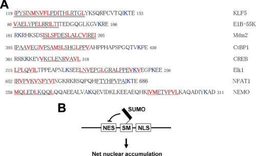
SUMOylation is a form of post-translational modification shown to control nuclear transport. Krüppel-like factor 5 (KLF5) is an important mediator of cell proliferation and is primarily localized to the nucleus. Here we show that mouse KLF5 is SUMOylated at lysine residues 151 and 202. Mutation of these two lysines or two conserved nearby glutamates results in the loss of SUMOylation and increased cytoplasmic distribution of KLF5, suggesting that SUMOylation enhances nuclear localization of KLF5. Lysine 151 is adjacent to a nuclear export signal (NES) that resembles a consensus NES. The NES in KLF5 directs a fused green fluorescence protein to the cytoplasm, binds the nuclear export receptor CRM1, and is inhibited by leptomycin and site-directed mutagenesis. SUMOylation facilitates nuclear localization of KLF5 by inhibiting this NES activity, and enhances the ability of KLF5 to stimulate anchorage-independent growth of HCT116 colon cancer cells. A survey of proteins whose nuclear localization is regulated by SUMOylation reveals that SUMOylation sites are frequently located in close proximity to NESs. A relatively common mechanism for SUMOylation to regulate nucleocytoplasmic transport may lie in the interplay between neighboring NES and SUMOylation motifs.


Figures

FIGURE 1.
FIGURE 1.
SUMOylation motifs in KLF5. A, a schematic showing the two ΨKXE SUMOylation motifs in KLF5. Shown also is the relative location of the zinc finger DNA binding domain of KLF5. B, conservation of the ΨKXE SUMOylation motifs in KLF5 from various species. The two ΨKXE motifs are located at residues 150-153 and 201-204 of mouse KLF5 (underlined).
FIGURE 2.
FIGURE 2.
SUMOylation of KLF5. A, SUMOylation of KLF5 with GFP-tagged SUMO-1 and identification of the two SUMOylation lysine residues within the ΨKXE motifs. COS-1 cells were co-transfected with GFP-SUMO-1 and one of the following: pMT3/HA-KLF5 (WT), pMT3/HA-KLF5-K151R (K151R), pMT3/HA-KLF5-K202R (K202R), or pMT3/HA-KLF5-K151R/K202R (K151R/K202R). Lysates were immunoprecipitated with a rabbit HA antibody followed by Western blotting with a mouse HA (lanes 1-4) or GFP (lanes 5-8) antibody. B, SUMOylation of KLF5 with His-tagged SUMO-1 and confirmation of the two SUMOylation sites. COS-1 cells were co-transfected with HIS-SUMO-1 and pMT3/HA-KLF5 (WT), pMT3/HA-KLF5-K151R (K151R), pMT3/HA-KLF5-K202R (K202R), or pMT3/HA-KLF5-K151R/K202R (K151R/K202R). The transfected cells were disrupted by boiling in the presence of SDS and NEM and Western blot was performed with a mouse HA, His, or β-actin antibody. C, SUMOylation of endogenous KLF5. Endogenous KLF5 is SUMOylated in the presence (lane 1, HS1) or absence (lane 2) of HIS-SUMO-1 transfection. D, loss in KLF5 SUMOylation by mutation at the two essential glutamate residues within the ΨKXE motifs. COS-1 cells were co-transfected with GFP-SUMO-1 and pMT3/HA-KLF5 (WT), pMT3/HA-KLF5-E153A (E153A), or pMT3/HA-KLF5-E153A/E204A (E153A/E204A). Lysates were immunoprecipitated with rabbit anti-HA followed by Western blot with mouse anti-HA (lanes 1-4) or GFP (lanes 5-8). A short exposure of the panel in lanes 5-8 is also shown. In all panels, * represents the di-SUMOylated form of KLF5; + and ++, mono-SUMOylated forms.
FIGURE 3.
FIGURE 3.
KLF5 is localized to both the nucleus and cytoplasm. A, localization of endogenous KLF5 to the cytoplasm in COS-1 cells. Cells were stained with a rabbit KLF5 antibody and RRX-conjugated donkey anti-rabbit secondary antibody. Cells were also stained with Hoechst dye to reveal the nuclei. The arrow shows punctate staining of KLF5 in the cytoplasm. B, immunostaining of KLF5 in DLD-1 cells. DLD-1 cells were transfected with siRNA against KLF5 or nonspecific control siRNA and subjected to immunofluorescence microscopy using a rabbit KLF5 antibody and Alexa Fluor 488-conjugated goat anti-rabbit secondary antibody. The brackets indicate specific punctate localization of KLF5 in the cytoplasm. C, subcellular fractionation of KLF5 in COS-1 cells. Cell lysates were fractionated into nuclear, cytosolic, and membrane fractions. An equal amount of each fraction was subjected to Western blot analysis with antibodies against KLF5, histone, tubulin, and Na+/K+-ATPase. D, Western blot analysis of KLF5 in DLD-1 cells following transfection with KLF5 siRNA or control siRNA. Cell lysates were divided into cytosolic and nuclear fractions and blotted against KLF5, histone, or actin antibodies.
FIGURE 4.
FIGURE 4.
Localization of KLF5 to vesicle-like structures in the cytoplasm. A, COS-1 cells were transfected with HA-KLF5 and immunostained with a rabbit HA antibody followed by RRX-conjugated donkey anti-rabbit secondary antibody. Hoechst stain was used to reveal the nuclei. The arrow indicates cytoplasmic staining in some of the cells expressing HA-KLF5. B, localization of HA-KLF5 to vesicle-like structures within the cytoplasm. Two cells exhibiting either largely nuclear distribution (top panels) or extensive cytoplasmic localization (bottom panels) of HA-KLF5 are shown. Progressively higher magnifications of a selected field are shown. Staining of the specific circular structures is indicated by arrowheads.
FIGURE 5.
FIGURE 5.
Co-localization of KLF5 and SNX3 in the cytoplasm. COS-1 cells were stained with rabbit KLF5 and goat SNX3 primary antibodies, followed by fluorescein isothiocyanate-conjugated donkey anti-rabbit (green) and RRX-conjugated donkey anti-goat (red) secondary antibodies, respectively. Cells were also stained with Hoechst dye to reveal the nuclei. Two cells with substantial cytoplasmic localization of KLF5 (A and B) and one cell with primarily nuclear KLF5 (C) are shown.
FIGURE 6.
FIGURE 6.
Demonstration of nucleocytoplasmic shuttling of KLF5 by heterokaryon assays. A, COS-1 cells were transfected with HA-KLF5 and fused with NIH3T3 cells in the presence of cycloheximide. Following fusion, cells were fixed and stained with a chicken HA antibody and fluorescein isothiocyanate-conjugated donkey anti-chicken secondary antibody. Nuclei were stained with Hoechst dye. Nuclei of murine cells were identified by their distinctive punctuate pattern (arrows). B, cells were treated under identical conditions but without fusion served as control.
FIGURE 7.
FIGURE 7.
SUMOylation facilitates nuclear localization of KLF5. COS-1 cells were transfected with pMT3/HA-KLF5 (WT), pMT3/HA-KLF5-K151R/K202R (K151R/K202R), or PMT3/HA-KLF5-E153A/E204A (E153A/E204A). Twenty-four h following transfection, cells were fixed and stained with a rabbit HA antibody and RRX-conjugated donkey anti-rabbit secondary antibody. A, images of two representative stained cells are shown for each construct. B, quantification of the percentage of cells exhibiting cytoplasmic staining. Shown are the averages and standard deviations of four independent experiments in which 100 cells were examined per experiment. *, p < 0.05 by two-tailed t test when compared with wild type (WT).
FIGURE 8.
FIGURE 8.
LRSs near the SUMOylation motifs of KLF5 and their GFP fusion constructs. Alignment of LRS1 (A) and LRS2 (B) from different species. The large hydrophobic residues (Φ) that match the NES consensus are indicated in red. The two SUMOylation motifs (SM) are underlined. Note that LRS2 lacks a conserved isoleucine (Ile207) that is critical for the NES consensus, whereas LRS1 completely matches the consensus regarding both the hydrophobic and spacing requirements as indicated by the top arrow (L/IPYSINMNVFL). Additional potential NESs that resemble the consensus sequence are shown by the two bottom arrows. C, GFP constructs linked to LRS1 or LRS2 and/or their adjacent SUMOylation motifs, SM1 or SM2, respectively. The mutated valine and leucine residues essential for the activity of LRS1 are underlined and the lysine and glutamate residues essential for KLF5 SUMOylation are also indicated (* and +).
FIGURE 9.
FIGURE 9.
LRS1 functions as a NES and is inhibited by SUMOylation. A, the construct containing GFP, GFP-LRS1, GFP-LRS2, or GFP-LRS1-VALA (GFP-LRS1-V127A/L129A), which contains mutation at the two residues predicted to be a critical part of the NES, was transfected into COS-1 cells, which were then visualized with an inverted fluorescence microscope. Shown are representative fluorescence images of transfected cells. B, COS-1 cells were transfected with the indicated GFP-LRS1-SM1, GFP-LRS1-SM1-K151R, or GFP-LRS1-SM1-E153A, and visualized with a fluorescence microscope. C, cells were transfected with the various constructs. Twenty-four h following transfection, cells were treated or not with 10 ng/ml leptomycin A and B for an additional 16 h. The percentages of cells with primarily cytoplasmic localization were tabulated. Shown are the averages and standard deviations of four independent experiments in which over 250 cells were examined per experiment. *, p < 0.05; **, p < 0.01; ***, p < 0.001 by two-tailed t test.
FIGURE 10.
FIGURE 10.
Inactivation of LRS1 by site-directed mutagenesis and inhibition of the cytoplasmic localization of KLF5 by leptomycin treatment. A, a schematic showing the various mutant constructs in the context of HA-KLF5. WT is wild type. VALA contains two point mutations substituting alanines for two residues within LRS1 predicated to be essential for NES activity (Val127 and Leu129). VALA/KRKR and VALA/EAEA are two constructs containing the NES mutation (V127A/L129A) and the two SUMOylation mutations (K151R/K202R and E153A/E204A). Numbers indicate the relative position of the residues mutated, which are marked with X. B, representative images of immunofluorescence studies of COS-1 cells transfected with the indicated constructs. LM is leptomycin. C, quantification of percentages of cells transfected with the various constructs that exhibit significant cytoplasmic staining as revealed by immunofluorescence. Shown are the averages and standard deviations of three independent experiments in which 100 cells were examined in each experiment. *, p < 0.05; **, p < 0.01 by two-tailed t test when compared with WT.
FIGURE 11.
FIGURE 11.
Interaction of CRM1 with LRS1. A, CRM1 binds to LRS1. COS-1 cells were transfected with the indicated GFP constructs. Forty-eight h following transfection, lysates were immunoprecipitated with a mouse CRM1 antibody. Western blotting was then conducted on the immunoprecipitates using goat GFP or mouse CRM1 antibodies. B, LRS1 but not LRS2 interacts with CRM1. Lysates from COS-1 cells transfected with the indicated constructs were immunoprecipitated with a rabbit GFP antibody and probed with goat GFP or mouse CRM1 antibodies. Both long and short exposures of the GFP images in the IP panel are provided to demonstrate SUMOylation of GFP-LRS1-SM1. C, endogenous SUMOylation of GFP-LRS1-SM1. Whole lysates from COS-1 cells transfected with the indicated constructs were subjected to Western blotting with a GFP antibody. The lower panel is a short exposure of the image in the upper panel. D, in vitro SUMOylation and CRM1 binding assay. The GFP fusion proteins were purified by immunoprecipitation and in vitro SUMOylated, followed by incubation with purified Ran/GTPγS and CRM1 proteins, as described under “Experimental Procedures.” The protein complexes were subjected to Western blotting with GFP and CRM1 antibodies. The asterisk indicates SUMOylated GFP-LRS1-SM1.
FIGURE 12.
FIGURE 12.
SUMOylation enhances the ability of KLF5 to stimulate anchorage-independent growth of HCT116 colorectal cancer cells. HCT116 cells were transfected with vector alone (V), pMT3/HA-KLF5 (WT), pMT3/HA-KLF5-K151R/K202R (K151R/K202R), or pMT3/HA-KLF5-E153A/E204A (E153A/E204A) and colony formation assay in soft agar conducted as previously described (43). Colonies were counted 3 weeks following transfection. A, a representative image of the colonies formed. B, quantitative results of the averages and standard deviations of four independent experiments. *, p < 0.05; **, p < 0.01; and ***, p < 0.001 by two-tailed t test.
FIGURE 13.
FIGURE 13.
Summary of regulation of nucleocytoplasmic transport by SUMOylation. A, alignment of various proteins of which nuclear localizations are facilitated by SUMOylation. The known SUMOylation sites that are involved in the regulation of nuclear transport are bolded in blue. The known NES motifs are double underlined and NES consensus sequences are underlined. The leucine or other large hydrophobic residues matching the NES consensus are bolded in red. Numbers are amino acid residue numbers. B, a model for the regulation of nucleocytoplasmic transport by SUMOylation. In this model, SUMOylation inhibits NES adjacent to a SUMOylation site, resulting in a net accumulation of the SUMOylated protein within the nucleus.

Similar articles

Cited by

References

    1. Johnson, E. S. (2004) Annu. Rev. Biochem. 73 355-382 - PubMed
    1. Wilson, V. G., and Rangasamy, D. (2001) Exp. Cell Res. 271 57-65 - PubMed
    1. Pichler, A., and Melchior, F. (2002) Traffic 3 381-387 - PubMed
    1. Terry, L. J., Shows, E. B., and Wente, S. R. (2007) Science 318 1412-1416 - PubMed
    1. Pemberton, L. F., and Paschal, B. M. (2005) Traffic 6 187-198 - PubMed

Publication types

MeSH terms