. 2008 Nov 7;283(45):31107-15.
doi: 10.1074/jbc.M803332200. Epub 2008 Sep 9.

Targeting of hFis1 to peroxisomes is mediated by Pex19p

Affiliations

Targeting of hFis1 to peroxisomes is mediated by Pex19p

Hannah K Delille et al. J Biol Chem. .

Abstract

The processes of peroxisome formation and proliferation are still a matter of debate. We have previously shown that peroxisomes share some components of their division machinery with mitochondria. hFis1, a tail-anchored membrane protein, regulates the membrane fission of both organelles by DLP1/Drp1 recruitment, but nothing is known about the mechanisms of the dual targeting of hFis1. Here we demonstrate for the first time that peroxisomal targeting of hFis1 depends on Pex19p, a peroxisomal membrane protein import factor. hFis1/Pex19p binding was demonstrated by expression and co-immunoprecipitation studies. Using mutated versions of hFis1 an essential binding region for Pex19p was located within the last 26 C-terminal amino acids of hFis1, which are required for proper targeting to both mitochondria and peroxisomes. The basic amino acids in the very C terminus are not essential for Pex19p binding and peroxisomal targeting, but are instead required for mitochondrial targeting. Silencing of Pex19p by small interference RNA reduced the targeting of hFis1 to peroxisomes, but not to mitochondria. In contrast, overexpression of Pex19p alone was not sufficient to shift the targeting of hFis1 to peroxisomes. Our findings indicate that targeting of hFis1 to peroxisomes and mitochondria are independent events and support a direct, Pex19p-dependent targeting of peroxisomal tail-anchored proteins.


Figures

FIGURE 1.
FIGURE 1.
Overview of the hFis1 constructs used in this study. The bars on the left side represent the proportion of the protein domains of the different tagged and/or truncated hFis1 constructs. Myc-hFis1 and GFP-hFis1 are full-length hFis1 constructs N-terminally fused with a Myc epitope tag or GFP, respectively. Myc-hFis1-ΔC has a C-terminal truncation of 5 amino acids whereas the Myc-hFis1-ΔTM/C construct lacks 26 amino acids (the transmembrane domain (TM) and the C terminus). In hFis1-YFP-TM/C these 26 amino acids are fused to the C terminus of YFP. GFP-hFis1K149/151A is a full-length construct carrying lysine to alanine mutations in the residues 149 and 151 (arrowheads). An overview of the particular binding to Pex19p and the subcellular localization is given on the right. ER, endoplasmic reticulum; Mito, mitochondria; and PO, peroxisomes.
FIGURE 2.
FIGURE 2.
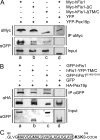
Pex19p and hFis1 interact with each other. A, COS-7 cells were co-transfected with Myc-hFis1 and YFP-Pex19p (on the left) or cytosolic YFP as negative control (pEYFP-N1; on the right), and incubated for 48 h. Whole cells were subjected to chemical cross-linking by adding DSP for 45 min. Immunoprecipitations (IPs) were performed with anti-GFP/YFP antibodies and Protein A-Sepharose followed by analysis of the samples by SDS-PAGE and immunoblotting using anti-Myc and anti-GFP/YFP antibodies. Note that Myc-hFis1 co-precipitated with YFP-Pex19p, but not with cytosolic YFP. The lower panel shows a positive control experiment without DSP. Pex11pβ-Myc, which is known to interact with Pex19p, forms a complex with YFP-Pex19p. B, COS-7 cells were transfected with Myc-hFis1 or not transfected. Immunoprecipitation of endogenous (and overexpressed) hFis1 was performed using anti-Fis1 antibodies and Protein A-Sepharose, and the co-precipitation of endogenous Pex19p was detected by immunoblotting using anti-Pex19 antibodies.
FIGURE 3.
FIGURE 3.
The Pex19p binding region is located in the C terminus of hFis1 and does not require basic amino acids. A, co-transfection of COS-7 cells with YFP-Pex19p and Myc-hFis1 (lane a), Myc-hFis1-ΔC(lane b), or Myc-hFis1-ΔTM/C (lane c) followed by immunoprecipitation (IP) with anti-Myc antibodies conjugated to agarose beads. The analysis of the samples was performed by immunoblotting using anti-GFP/YFP antibodies. Note that Pex19p binding to hFis1 is impaired after removal of the C-terminal tail or the transmembrane domain of hFis1 (lanes b and c). To exclude unspecific co-precipitations control experiments were performed with cytosolic YFP (pEYFP-N1) and Myc-hFis1 (lane d). B, COS-7 cells were co-transfected with HA-Pex19p and GFP-hFis1 (lane a), hFis1-YFP-TM/C (lane b), GFP-hFis1K149/151A (lane c), or cytosolic GFP as negative control (pEGFP-N1; lane d). IPs were performed by adding anti-GFP/YFP antibodies and Protein A-Sepharose. The samples were analyzed by immunoblotting with anti-HA antibodies. Note that the C-terminal domain of hFis1 (TM/C, 26 amino acids) fused to YFP is sufficient to co-precipitate HA-Pex19p (lane b) and that mutations of the basic amino acids in the very C terminus of hFis1 do not abolish binding to HA-Pex19p (lane c). C, amino acid sequence of the C terminus (amino acids 122-152) of hFis1. The transmembrane domain is boxed, and basic amino acids (Lys-149 and Lys-151) are in bold. Quantitative analyses of A and B are shown in supplemental Fig. S1.
FIGURE 4.
FIGURE 4.
C-terminal basic amino acids are not required for peroxisomal targeting of hFis1. COS-7 cells expressing GFP-hFis1K149/151A (D-L) or GFP-hFis1 (A-C) were processed for immunofluorescence using anti-PMP70 antibodies to label peroxisomes (H and K) or anti-Tom20 antibodies to label mitochondria (B and E). G-I, overview of a representative cell. J-L, higher magnification view. Arrows highlight some regions of colocalization. Overlays (merge) are shown on the right. Note that GFP-hFis1K149/151A is still prominently targeted to peroxisomes, whereas staining of mitochondria is weak. In addition, GFP-hFis1K149/151A is misdirected to the tubulo-reticular network of the ER. Bars, 10 μm. See also supplemental Fig. S2 for a peroxisomal co-labeling of cells expressing GFP-hFis1.
FIGURE 5.
FIGURE 5.
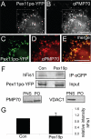
Overexpression of Pex19p does not increase peroxisomal targeting of hFis1. A-E, Pex11pα-YFP is properly targeted to peroxisomes. COS-7 cells expressing Pex11pα-YFP were immunostained with an antibody to PMP70 (B and D). C-E, higher magnification view of the boxed regions in A and B. E, overlay (merge) of C and D. F, peroxisome immunoprecipitation. COS-7 cells were transfected with Pex11pα-YFP alone (Con) or co-transfected with HA-Pex19p. Peroxisomal membranes were immunoprecipitated from peroxisome-enriched fractions using anti-GFP/YFP antibodies and Protein A-Sepharose. Endogenous hFis1 was detected by immunoblotting with anti-hFis1 antibodies. Contamination of mitochondria in the peroxisomal membrane fractions (PO) was low, as shown by immunoblotting with VDAC1 antibodies in the lower panel, in contrast to the enrichment of peroxisomal markers (PMP70). Equal amounts of protein (30 μg/lane) were loaded onto the gels. G, quantitative analysis of the anti-hFis1 immunoblots. Data were normalized using Pex11pα as input references. The data are from four independent experiments and are expressed as means ± S.D. N, nucleus. PNS, postnuclear supernatant. Bars, 10 μm.
FIGURE 6.
FIGURE 6.
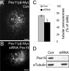
Silencing of Pex19p reduces peroxisomal proliferation induced by Pex11pβ. COS-7 cells were transfected with Pex19p siRNA duplexes. After 48 h the cells were transfected with Pex11pβ-Myc and after additional 6 h processed for immunofluorescence using anti-Myc (and anti-PMP70) antibodies. A, control cell (Con) with typical tubular, elongated peroxisomes induced by Pex11pβ expression. B, silencing of Pex19p (siRNA). In many cases, the peroxisomes exhibit a spherical morphology and are not elongated. C, quantitative evaluation of peroxisome morphology. Note the reduced frequency of tubular peroxisomes in cells silenced for Pex19p. The data are from four independent experiments and are expressed as means ± S.D. (**, p < 0.01). D, immunoblots of cell lysates prepared after 48 h from control (Con) and silenced cells (siRNA) using anti-Pex19p and anti-tubulin antibodies (loading control). Equal amounts of protein (35 μg/lane) were loaded onto the gels. N, nucleus. Bars, 10 μm.
FIGURE 7.
FIGURE 7.
Silencing of Pex19p inhibits the peroxisomal targeting of hFis1. Pex19p in COS-7 cells was silenced by transfection with siRNA duplexes (D-I). After 48 h the cells were transfected with GFP-hFis1, and the targeting of GFP-hFis1 to peroxisomes was assayed after 3 h. Peroxisomes were immunostained with antibodies to PMP70 (B, E, and H). Co-localization of GFP-hFis1 and peroxisomes in control cells (A-C), and in cells silenced for Pex19p (D-I). Overlays (merge) are shown on the right. Arrows in (A-C) highlight regions of co-localization. Note the absence or reduction of co-localization (arrows) of GFP-hFis1 with PMP70 after silencing of Pex19p (D-I). J, fluorescence intensities of GFP-hFis and PMP-TRITC were determined on digital images by highlighting (encircling) single peroxisomes, and the GFP/TRITC ratios were calculated. 60-140 peroxisomes per cell were analyzed in ∼60 control as well as treated cells. Data are expressed as means ± S.D. (**, p < 0.01) and are from three independent experiments. N, nucleus. Bars, 10 μm.
FIGURE 8.
FIGURE 8.
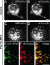
Silencing of Pex19p does not inhibit mitochondrial targeting of hFis1. COS-7 cells were processed as described in Fig. 7. Mitochondria were immunostained with an antibody to hTom22c, a mitochondrial outer membrane protein. A and B, control cells. C-G, COS-7 cells transfected with siRNA specific for Pex19p. E-G, higher magnification view. G, overlay (merge) of E and F. N, nucleus. Bars, 10 μm.

Similar articles

Cited by

References

    1. Schrader, M., and Yoon, Y. (2007) BioEssays 29 1105-1114 - PubMed
    1. Schrader, M. (2006) Biochim. Biophys. Acta 1763 531-541 - PubMed
    1. Smirnova, E., Griparic, L., Shurland, D. L., and van der Bliek, A. M. (2001) Mol. Biol. Cell 12 2245-2256 - PMC - PubMed
    1. Yoon, Y., Krueger, E. W., Oswald, B. J., and McNiven, M. A. (2003) Mol. Cell. Biol. 23 5409-5420 - PMC - PubMed
    1. Koch, A., Thiemann, M., Grabenbauer, M., Yoon, Y., McNiven, M. A., and Schrader, M. (2003) J. Biol. Chem. 278 8597-8605 - PubMed

Publication types