. 2008 Nov 14;283(46):31968-80.
doi: 10.1074/jbc.M806050200. Epub 2008 Sep 9.

Obscurin targets ankyrin-B and protein phosphatase 2A to the cardiac M-line

Affiliations

Obscurin targets ankyrin-B and protein phosphatase 2A to the cardiac M-line

Shane R Cunha et al. J Biol Chem. .

Abstract

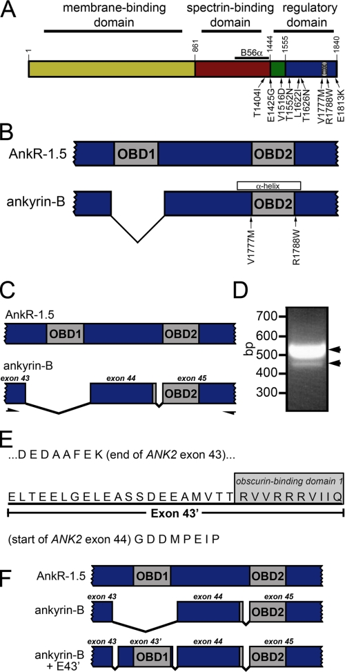
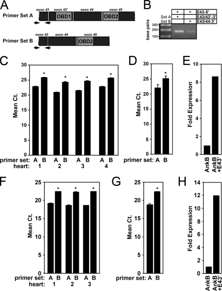
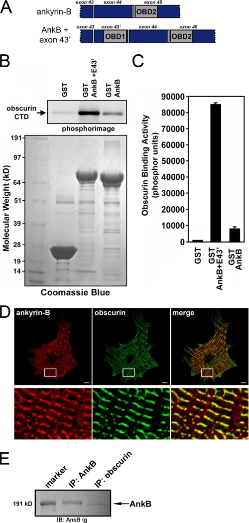
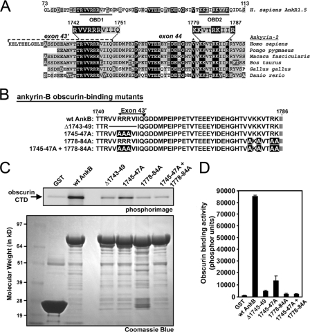
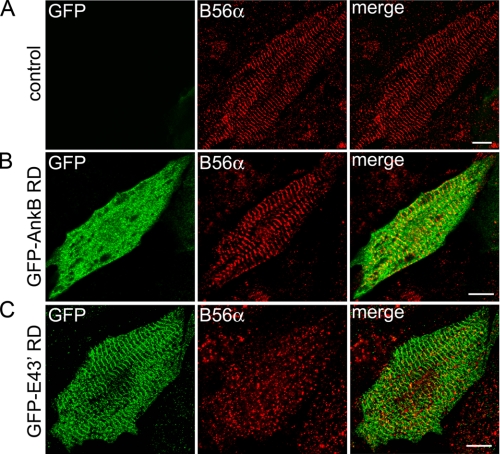
Ankyrin-B targets ion channels and transporters in excitable cells. Dysfunction in ankyrin-B-based pathways results in defects in cardiac physiology. Despite a wealth of knowledge regarding the role of ankyrin-B for cardiac function, little is known regarding the mechanisms underlying ankyrin-B regulation. Moreover, the pathways underlying ankyrin-B targeting in heart are unclear. We report that alternative splicing regulates ankyrin-B localization and function in cardiomyocytes. Specifically, we identify a novel exon (exon 43') in the ankyrin-B regulatory domain that mediates interaction with the Rho-GEF obscurin. Ankyrin-B transcripts harboring exon 43' represent the primary cardiac isoform in human and mouse. We demonstrate that ankyrin-B and obscurin are co-localized at the M-line of myocytes and co-immunoprecipitate from heart. We define the structural requirements for ankyrin-B/obscurin interaction to two motifs in the ankyrin-B regulatory domain and demonstrate that both are critical for obscurin/ankyrin-B interaction. In addition, we demonstrate that interaction with obscurin is required for ankyrin-B M-line targeting. Specifically, both obscurin-binding motifs are required for the M-line targeting of a GFP-ankyrin-B regulatory domain. Moreover, this construct acts as a dominant-negative by competing with endogenous ankyrin-B for obscurin-binding at the M-line, thus providing a powerful new tool to evaluate the function of obscurin/ankyrin-B interactions. With this new tool, we demonstrate that the obscurin/ankyrin-B interaction is critical for recruitment of PP2A to the cardiac M-line. Together, these data provide the first evidence for the molecular basis of ankyrin-B and PP2A targeting and function at the cardiac M-line. Finally, we report that ankyrin-B R1788W is localized adjacent to the ankyrin-B obscurin-binding motif and increases binding activity for obscurin. In summary, our new findings demonstrate that ANK2 is subject to alternative splicing that gives rise to unique polypeptides with diverse roles in cardiac function.


Figures

FIGURE 1.
FIGURE 1.
Novel ANK2 exon encodes putative obscurin-binding domain. A, ankyrin domain organization. Ankyrin-B contains three major domains, including the membrane-binding domain (yellow), spectrin-binding domain (red), and regulatory domain. The RD is comprised of the death domain (green) and C-terminal domain (blue). Identified ankyrin-B loss-of-function variants associated with human arrhythmia are noted on the diagram. Previously identified amphipathic α-helix in RD (28) is indicated with circles. B56α-binding site in the spectrin-binding domain is represented by a black line. B, both obscurin-binding domains (OBD1 and OBD2) in AnkR1.5 are required for AnkR/obscurin interaction. Ankyrin-B lacks OBD1. Ankyrin-B variants in this region are noted on the diagram. C, PCR-based strategy to identify novel ANK2 exon(s) encoding OBD1 in ankyrin-B. PCR primers were designed against ANK2 sequence in exons 43 and 45 to amplify cardiac ankyrin-B transcripts. D, ethidium bromide agarose gel demonstrates amplification of two PCR products (518 and 425 bp) from human heart cDNA. In the 518-bp PCR product, the additional 93 bp represent a new, previously unreported exon (exon 43′). E, exon 43′ encodes 31 amino acids that contain a homologous site to OBD1 in the terminal 10 amino acids. F, with the addition of exon 43′, human ankyrin-B + E43′ contains two homologous sites corresponding to OBD1 and OBD2 in small AnkR-1.5.
FIGURE 2.
FIGURE 2.
Ankyrin-B + E43is the primary ANK2 transcript in vertebrate heart. A, location of exon-exon spanning primers to PCR amplify ANK2 transcripts + exon 43′ (primer set A) and ANK2 transcriptsexon 43′ (primer set B). B, ethidium bromide-agarose gel demonstrating PCR amplification of ANK2 transcripts from mRNA isolated from human left ventricular tissue in the presence of the full-length exon-exon spanning primers (primer set A, 1st column; primer set B, 2nd column). In contrast, no PCR products are detected using half the boundary-spanning primer (data not shown). C, relative mRNA expression in four separate human hearts of ANK2 transcripts + exon 43′ (primer set A) and ANK2 transcriptsexon 43′ (primer set B) expressed as mean cycle threshold (Ct) values. D, average of the Ct values from four hearts (p < 0.05). E, fold expression for mRNA transcripts of human ANK2 ± exon 43′. Note that transcripts containing exon 43′ are expressed ∼8.5-fold greater than transcripts lacking exon 43′. F-H, relative mRNA expression in three mouse hearts of Ank2 ± exon 43′. Note that transcripts containing exon 43′ are expressed 12-fold greater than transcripts lacking exon 43′. Error bars represent standard deviations (n = 3, p < 0.05).
FIGURE 3.
FIGURE 3.
Binding and co-localization of ankyrin-B with obscurin; an association that requires novel obscurin-binding domain 1. A, organization for ankyrin-B RD constructs ± exon 43′. B, enhanced obscurin binding activity for ankyrin-B construct containing both OBD1 and -2. Upper panel, phosphorimage of obscurin CTD labeled with [35S]methionine bound to GST fusion proteins of AnkB RD ± exon 43′. Lower panel, Coomassie Blue stain of the same protein gel that demonstrates equal loading of GST and GST fusion proteins. C, quantification of obscurin binding activity. Error bars represent standard deviations (n = 3). D, upper panels, immunofluorescent localization of ankyrin-B (red), obscurin (green), and their co-localization in neonatal mouse cardiomyocytes. Scale bar is 10 μm. Regions boxed in white are magnified in the lower panels. E, ankyrin-B immunoblot (IB) demonstrating co-immunoprecipitation (IP) of ankyrin-B from rat heart lysate with antibodies to ankyrin-B or obscurin.
FIGURE 4.
FIGURE 4.
Mutations to OBD1 and -2 disrupt ankyrin-B/obscurin interactions. A, alignment of ankyrin-B RD from various species and human small AnkR1.5. Accession numbers for species-specific ankyrin-B transcripts are Q01484 (Homo sapiens), CR773816 (Pongo pygmaeus), CJ448751 (Macaca fascicularis), DV930431 (Bos taurus), AL588116 (Gallus gallus), and EB895593 (Danio rerio). All of the non-human sequences are derived from translated genomic sequences and have been confirmed in expressed sequence tags. Background color corresponds to the level of amino acid homology: black, identical; light gray, conserved; and dark gray, similar. For AnkR1.5, the double-underlined regions represent obscurin-binding domains 1 and 2 (21). The location of ANK2 exon 43′ is indicated in H. sapiens ankyrin-B. Asterisks denote location of ankyrin-B variants associated with human arrhythmia. B, diagram of ankyrin-B RD obscurin-binding mutants. C, mutations to specific residues in ankyrin-B OBD1 and -2 disrupt ankyrin-B/obscurin interactions. Upper panel, phosphorimage of obscurin CTD labeled with [35S]methionine bound to GST fusion proteins of ankyrin-B RD mutants. Lower panel, Coomassie Blue stain of the corresponding protein gel that demonstrates equal loading of GST fusion proteins. D, quantification of obscurin binding activity (phosphor units). Error bars represent standard deviations (n = 3).
FIGURE 5.
FIGURE 5.
Ankyrin-B is not required for obscurin targeting in native cardiomyocytes. Subcellular localization of ankyrin-B and obscurin in ankyrin-B wild-type (A), ankyrin-B+/- (B), and ankyrin-B-/- cardiomyocytes (C). Scale bar represents 10 μm. Lower panels represent magnified views of the areas boxed in white. Note that loss of ankyrin-B does not affect the expression/localization of obscurin.
FIGURE 6.
FIGURE 6.
OBD1 regulates the targeting of ankyrin-B RD to the M-line. A, lentiviral expression of GFP-AnkB RD lacking exon 43′ that contains OBD1 is diffusely cytoplasmic in neonatal rat cardiomyocytes. Note the expression of this construct by immunofluorescent detection of GFP does not co-localize with obscurin (red) at M-lines (merge). B, lentiviral expression of GFP-AnkB RD with exon 43′ (GFP-E43RD) co-localizes with obscurin at M-lines (merge). Scale bar represents 10 μm.
FIGURE 7.
FIGURE 7.
Ankyrin-B RD with exon 43inhibits association of endogenous ankyrin-B with obscurin. A, immunolocalization of viral GFP-AnkB RD (lacking OBD1) with endogenous ankyrin-B. Expression of endogenous ankyrin-B was detected using the ankyrin-B mAb that was generated against SBD; therefore, this antibody will not recognize virally expressed GFP-AnkB RD. GFP-AnkB RD is cytoplasmic and does not co-localize with endogenous ankyrin-B at the M-line. B, immunolocalization of viral ankyrin-B containing OBD1 and -2 (GFP-E43RD) with endogenous ankyrin-B. Viral expression of GFP-E43′ RD decreases localization of endogenous ankyrin-B at the M-line. Scale bars represent 10 μm. C, immunoblot (IB) analysis of endogenous ankyrin-B associated with GST-obscurin CTD in the presence of competitors GFP-AnkB RD or GFP-E43′ RD. Heart lysate was incubated with a limited amount of GST-obscurin CTD and varying amounts of competitors (5, 10, and 20 μg). Immunoprecipitated (IP) complexes were probed for endogenous ankyrin-B (upper panels) and GFP (lower panels). Note that the level of endogenous ankyrin-B associated with obscurin CTD decreases in the presence of increasing amounts of GFP-E43′ RD (upper right panel) but not with GFP-AnkB RD (upper left panel). Correspondingly, there is an increase in GFP-E43′ RD associated with obscurin (lower right panel) as the amount of this competitor is increased (bottom right panel). In contrast, GFP-AnkB RD does not associate with obscurin (lower left panel, denoted by asterisk). Therefore, the association of endogenous ankyrin-B with obscurin is unaffected by GFP-AnkB RD (upper left panel).
FIGURE 8.
FIGURE 8.
Ankyrin-B is required for B56α M-line targeting in primary adult cardiomyocytes. Subcellular localization of ankyrin-B and PP2A regulatory subunit B56α in ankyrin-B wild-type (A and B) and ankyrin-B+/- (D and E) adult cardiomyocytes. Note that loss of ankyrin-B results in clear loss of M-line localization of B56α. C and F, α-actinin staining was used to confirm cell viability following Langendorff isolation of myocytes (note that loss of ankyrin-B does not affect α-actinin expression or localization). Scale bars represent 10 μm.
FIGURE 9.
FIGURE 9.
Ankyrin-B/obscurin interaction is required for B56α M-line targeting in primary cardiomyocytes. Control cardiomyocytes (A) or cardiomyocytes transduced with equal multiplicities of infection of GFP-ankyrin-B RD (B) or GFP-ankyrin-B RD + E43′ (GFP-E43′ RD) (C) were immunostained with GFP and B56α-specific Igs and analyzed by confocal microscopy. Note that M-line expression of ankyrin-B associated protein B56α is reduced by GFP-E43′ RD (C) but not by GFP-AnkB RD (B). Scale bars represent 10 μm.
FIGURE 10.
FIGURE 10.
Ankyrin-B human arrhythmia disease mutation R1788W increases ankyrin/obscurin binding activity. A, ankyrin-B human mutation R1788W associated with arrhythmia displays increased binding activity for obscurin when introduced into ankyrin-B RD. Ankyrin-B mutation V1777M associates with obscurin similar to wild-type ankyrin-B RD. Upper panel, phosphorimage of obscurin CTD labeled with [35S]methionine bound to GST fusion proteins of ankyrin-B RD containing the missense variants V1777M or R1788W. Lower panel, Coomassie Blue stain of the corresponding protein gel that demonstrates equal loading of GST fusion proteins. B, quantification of obscurin binding activity (phosphor units). Error bars represent standard deviation (n = 3, p < 0.05).

Similar articles

Cited by

References

    1. Scotland, P., Zhou, D., Benveniste, H., and Bennett, V. (1998) J. Cell Biol. 143 1305-1315 - PMC - PubMed
    1. Mohler, P. J., Schott, J. J., Gramolini, A. O., Dilly, K. W., Guatimosim, S., duBell, W. H., Song, L. S., Haurogne, K., Kyndt, F., Ali, M. E., Rogers, T. B., Lederer, W. J., Escande, D., Le Marec, H., and Bennett, V. (2003) Nature 421 634-639 - PubMed
    1. Mohler, P. J., Splawski, I., Napolitano, C., Bottelli, G., Sharpe, L., Timothy, K., Priori, S. G., Keating, M. T., and Bennett, V. (2004) Proc. Natl. Acad. Sci. U. S. A. 101 9137-9142 - PMC - PubMed
    1. Mohler, P. J., Le Scouarnec, S., Denjoy, I., Lowe, J. S., Guicheney, P., Caron, L., Driskell, I. M., Schott, J. J., Norris, K., Leenhardt, A., Kim, R. B., Escande, D., and Roden, D. M. (2007) Circulation 115 432-441 - PubMed
    1. Davis, J. Q., and Bennett, V. (1994) J. Biol. Chem. 269 27163-27166 - PubMed

Publication types

MeSH terms