. 2008 Oct;36(18):5787-99.
doi: 10.1093/nar/gkn576. Epub 2008 Sep 9.

The human mitochondrial ribosome recycling factor is essential for cell viability

Affiliations

The human mitochondrial ribosome recycling factor is essential for cell viability

Joanna Rorbach et al. Nucleic Acids Res. 2008 Oct.

Abstract

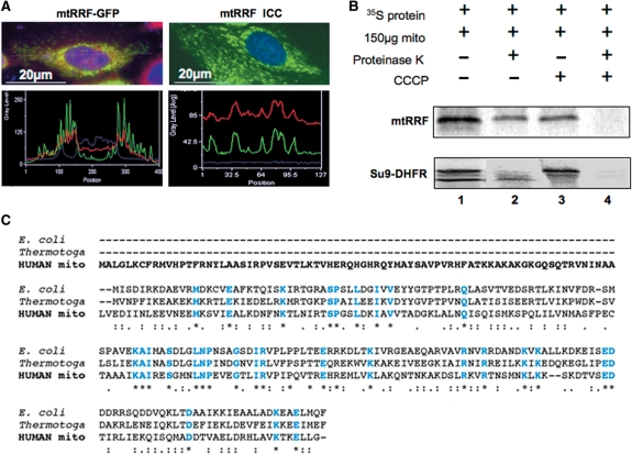
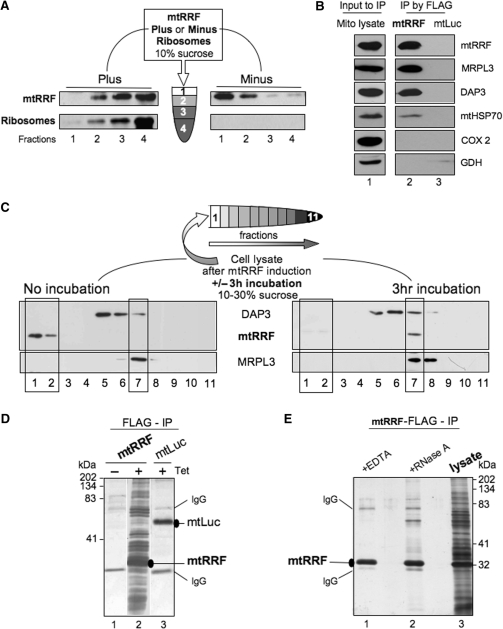
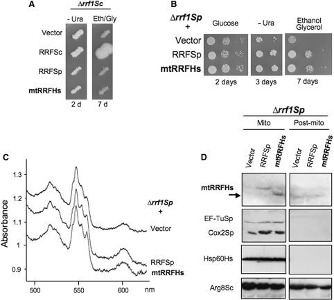
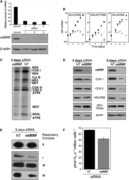
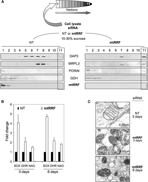
The molecular mechanism of human mitochondrial translation has yet to be fully described. We are particularly interested in understanding the process of translational termination and ribosome recycling in the mitochondrion. Several candidates have been implicated, for which subcellular localization and characterization have not been reported. Here, we show that the putative mitochondrial recycling factor, mtRRF, is indeed a mitochondrial protein. Expression of human mtRRF in fission yeast devoid of endogenous mitochondrial recycling factor suppresses the respiratory phenotype. Further, human mtRRF is able to associate with Escherichia coli ribosomes in vitro and can associate with mitoribosomes in vivo. Depletion of mtRRF in human cell lines is lethal, initially causing profound mitochondrial dysmorphism, aggregation of mitoribosomes, elevated mitochondrial superoxide production and eventual loss of OXPHOS complexes. Finally, mtRRF was shown to co-immunoprecipitate a large number of mitoribosomal proteins attached to other mitochondrial proteins, including putative members of the mitochondrial nucleoid.


Figures

Figure 1.
Figure 1.
mtRRF is a mitochondrial protein with an extended N-terminal pre-sequence. (A) Human mtRRF is targeted to mitochondria. Left panels show HeLa cells transiently transfected (24 h) with a mtRRF–GFP fusion construct. Cells were stained to visualize nuclei (DAPI blue) and mitochondria (Mitotracker red). Fluorescence images and linescans confirmed mitochondrial localization of mtRRF–GFP by superimposition of green and red signals (lower left). The image reflects three-independent transfections. Endogenous mtRRF in HeLa cells was visualized by immunocytochemistry (upper right) using affinity purified anti-mtRRF and FITC secondary. DAPI-stained nuclei (blue) and Mitotracker the mitochondria (red). Mitochondrial localization of mtRRF was confirmed by superimposition of linescan green and red fluorescence (lower right). The image reflects three-independent transfections. (B) Human mtRRF is imported into mitochondria. FL 35S-radiolabelled mtRRF was in vitro synthesized and incubated with rat liver mitochondria (lane 1). Under import conditions a single product is visible (lane 2) that is protected from proteinase K (lane 3), but degraded by treatment with proteinase K and FCCP uncoupler (lane 4). Control import reactions contained DHFR with a mitochondrial pre-sequence (Su9) showing the FL pre-protein and the matured form under import conditions. (C) Sequence alignment (CLUSTALW) indicates an extensive N-terminal pre-sequence when compared with E. coli or Thermotoga maritima. Identity to these RRFs is indicated in blue and by *, high levels of similarity by a colon ‘:’ and lower levels by a fullstop ‘.’.
Figure 2.
Figure 2.
Human mtRRF associates with bacterial ribosomes in vitro and mitoribosomes in vivo. (A) Human mtRRF binds E. coli ribosomes. Recombinant mtRRF (mtRRFΔ69) was incubated with or without 70S ribosomes prior to sedimentation through 10% (w:v) sucrose. Fractions were subjected to western analysis. (B) mtRRF associates with mitoribosomes. FLAG-mtRRF and mtLuc HEK293T cells were induced, mitochondria isolated. Lysate was prepared (lane 1) from which proteins were immunoprecipitated via the FLAG epitope (lanes 2–3). Western blot analysis used antibodies to mtRRF, large 39S and small 28S mitoribosomal subunits (MRPL3, DAP3), mitochondrial chaperone (mtHSP70), complex IV (COX 2) and a matrix protein (glutamate dehydrogenase -GDH). (C) Pre-incubation of mitochondria allows detection of mtRRF binding to mitoribosomes. Lysate was prepared from HEK293T cells after induction of FLAG-mtRRF and was either separated immediately or post 3 h incubation on a 10–30% (v:v) isokinetic sucrose gradient. Fractions were analysed by western blot with antibodies to mtRRF, MRPL3 (39S mitoribosomal subunit) or DAP3 (28S mitoribosomal subunit). A similar time-dependent mitoribosomal association in HeLa cell lysate was noted for the endogenous mtRRF (data not shown). (D) HEK293T cells were induced (±Tet) for FLAG-mtRRF (lanes 1–2) or control mtluc (lane 3) expression and mitochondria isolated. Co-immunoprecipitating proteins were separated through a 12% SDS–PAG and visualized by silver stain. mtRRF, mtluc and anti-FLAG IgG are indicated. (E) Preparations of the mtRRF IP were also treated with EDTA (lane 1) or RNase A (lane 2). Mitochondrial lysate is shown in lane 3.
Figure 3.
Figure 3.
Human mtRRF suppresses a partial respiratory deficiency in Δrrf1 fission yeast. (A) Human mtRRF cannot rescue the Δrrf respiratory defect in budding yeast. Saccharomyces cerevisiae transformants of Δrrf1 strain MG38 carrying the control vector pFL44L or URA3 plasmids producing the S. cerevisiae, S. pombe or human mtRRF were replica-plated on non-fermentable media (eth/gly + glycerol 0.1% glucose) and incubated at 28°C. (B–D) Human mtRRF restores respiratory capacity in Δrrf1 fission yeast. Schizosaccharomyces pombe lacking endogenous RRF (Δrrf1Sp, NB331) was transformed with vector alone (vector) or expressing human (RRFHs) or S. pombe (RRFSp) mtRRF (B). Serial dilutions of three Δrrf1Sp transformants were spotted on complete glucose, uracil-free minimal or complete gly/eth media and grown at 28 °C for the times indicated. (C) Δrrf1Sp transformants were grown on 2% glycerol/0.1% glucose (vector) or ethanol/glycerol medium (RRFSp or RRFHs) before recording whole-cell cytochrome spectra. Cytochrome b + c1 and aa3 correspond to Complex III and Complex IV, respectively. (D) Mitochondria and post-mitochondrial supernatants from transformants grown in glucose medium minus uracil were separated by 12% PAGE and analysed by western blot with antibodies recognizing human mtRRF, S. pombe mtEF-Tu and mt-encoded Cox2, human mt-Hsp60 and S. cerevisiae mtArg8 (this also recognizes S. pombe Arg1 in mitochondria and an unknown protein in the supernatant).
Figure 4.
Figure 4.
Depletion of human mtRRF severely affects cell viability and compromises mitochondrial translation. (A) siRNA-mediated depletion of human mtRRF. HeLa cells were exposed to three different siRNA molecules all in the mtRRF-coding sequence or an control siRNA for 6 days and RNA or cell lysates were prepared. Levels of mtRRF transcript compared to control siRNA transfected cells were quantified by real-time PCR (upper graph, mean ± SEM from four-independent transfections). Western blots (30 µg lysate/lane) in the lower panel were performed with antibodies against mtRRF to confirm depletion and β-actin as a loading. The blot accurately reflects three experiments. (B) Depletion of mtRRF causes a severe growth defect. Multiple aliquots of HeLa cells (left and centre panels) were exposed to targeted (mtRRF, open triangles) or non-targeted (NT, black squares) siRNA for 6 days in glucose (left) or galactose (centre) media, cells counted and presented in a semi-log plot. Counts were made of HEK293T transfectants (right panel) treated with non-targeted siRNA (NT, black squares) or siRNA directed against the endogenous mtRRF 3′-UTR with (open circles) or without (open triangles) concomitant inducible expression of FLAG-mtRRF. Numbers are a mean ± SEM of four independent wells. (C) In vivo mitochondrial proteins synthesis is partially affected by mtRRF depletion. HeLa cells were grown in the presence of control (NT) or targeted (mtRRF) siRNA. Cytosolic protein synthesis was inhibited with emetine prior to labelling mitochondrial proteins with 35S-methionine (15 min). Equal amounts of lysate (20 µg) were separated by 15% SDS–PAGE and gels analysed by PhosphorImager. Polypeptides designation is as described in ref. (23). (D) Steady-state levels of mtDNA-encoded proteins are affected by depletion of mtRRF. Western blots show analysis of cell lysates after 3 or 6 days siRNA treatment (mtRRF or NT) with antibodies against mitochondrial translation products or proteins sensitive to mitochondrial translation inhibition (cytochrome c oxidase subunits I and II—COX I and 2; complex I subunit NDUFB8) or nuclear-encoded complex II SDH 70 kDa protein and β-actin as a loading control. The blot accurately reflects three repeat experiments. (E) OXPHOS complexes are reduced at steady-state levels after mtRRF depletion. HeLa cells were exposed (6 days) to mtRRF- or NT-targeted siRNA prior to BN–PAGE. Complexes were visualized with antisera to complex I, anti-39 kDa; complex II, anti-SDH 70 kDa; complex III, anti-core 2, complex IV, anti-COII. The blot accurately reflects three experiments. SDH is a complex only comprising proteins encoded in the cytosol. (F) Respiratory coupling is modestly affected in cells depleted of human mtRRF. HeLa cells were exposed (3 days) to mtRRF-or NT-targeted siRNA prior to high resolution respirometry (as in Methods), three measures of respiratory control and capacity were made.
Figure 5.
Figure 5.
Reduction of mtRRF results in redistribution of ribosomal proteins, increased ROS and mitochondrial dysmorphism. (A). HeLa lysates were prepared from cells treated with mtRRF or non-targeted (NT) siRNA (3 days). After separation through 10–30% sucrose gradients, fractions were analysed by western blot using antibodies against the small (DAP3) and large (MRPL3) mitochondrial ribosomal subunits, mtRRF, porin as a mitochondrial membrane marker and glutamate dehydrogenase (GDH, matrix). (B) Superoxide levels are increased after mtRRF depletion. HeLa cells were exposed to mtRRF (white) or non-targeted (NT, black) siRNA over 6 days and superoxide and peroxide levels were measured with mitoSOX (superoxide) or DHR (peroxide) and compared to untreated controls. The fold increase is shown as a mean ± SEM from minimally three repeat experiments. Mitochondrial mass per cell was also measured using the cardiolipin selective dye NAO. (C) HeLa cells depleted of mtRRF show altered morphology. EM micrographs show HeLa cells treated with mtRRF (3 and 6 days) or non-targeted (NT, 3 days) siRNA. The images reflect mitochondria from two preparations.

Similar articles

Cited by

References

    1. Anderson S, Bankier AT, Barrell BG, De Bruijn MHL, Coulson AR, Drouin J, Eperon IC, Nierlich DP, Roe BA, Sanger F, et al. Sequence and organization of the human mitochondrial genome. Nature. 1981;290:457–465. - PubMed
    1. Pel HJ, Grivell LA. Protein synthesis in mitochondria. Mol. Biol. Rep. 1994;19:183–194. - PubMed
    1. Chung HK, Spremulli LL. Purification and characterization of elongation factor G from bovine liver mitochondria. J. Biol. Chem. 1990;265:21000–21004. - PubMed
    1. Koc EC, Spremulli LL. Identification of mammalian mitochondrial translational initiation factor 3 and examination of its role in initiation complex formation with natural mRNAs. J. Biol. Chem. 2002;277:35541–35549. - PubMed
    1. Schwartzbach CJ, Spremulli LL. Bovine mitochondrial protein synthesis elongation factors. Identification and initial characterization of an elongation factor Tu-elongation factor Ts complex. J. Biol. Chem. 1989;264:19125–19131. - PubMed

Publication types

MeSH terms

Substances