. 2008 Oct;36(18):5773-86.
doi: 10.1093/nar/gkn552. Epub 2008 Sep 9.

Loss of DNA ligase IV prevents recognition of DNA by double-strand break repair proteins XRCC4 and XLF

Affiliations

Loss of DNA ligase IV prevents recognition of DNA by double-strand break repair proteins XRCC4 and XLF

Sumithra Jayaram et al. Nucleic Acids Res. 2008 Oct.

Abstract

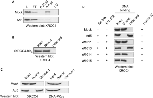
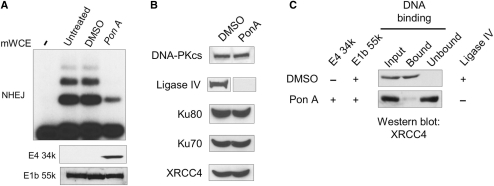
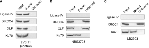
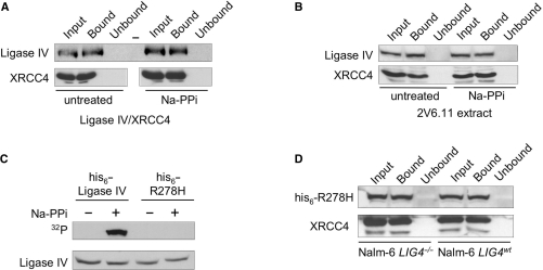
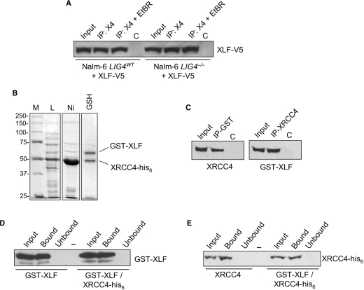
The repair of DNA double-strand breaks by nonhomologous end-joining (NHEJ) is essential for maintenance of genomic integrity and cell viability. Central to the molecular mechanism of NHEJ is DNA ligase IV/XRCC4/XLF complex, which rejoins the DNA. During adenovirus (Ad5) infection, ligase IV is targeted for degradation in a process that requires expression of the viral E1B 55k and E4 34k proteins while XRCC4 and XLF protein levels remain unchanged. We show that in Ad5-infected cells, loss of ligase IV is accompanied by loss of DNA binding by XRCC4. Expression of E1B 55k and E4 34k was sufficient to cause loss of ligase IV and loss of XRCC4 DNA binding. Using ligase IV mutant human cell lines, we determined that the absence of ligase IV, and not expression of viral proteins, coincided with inhibition of DNA binding by XRCC4. In ligase IV mutant human cell lines, DNA binding by XLF was also inhibited. Expression of both wild-type and adenylation-mutant ligase IV in ligase IV-deficient cells restored DNA binding by XRCC4. These data suggest that the intrinsic DNA-binding activities of XRCC4 and XLF may be subject to regulation and are down regulated in human cells that lack ligase IV.


Figures

Figure 1.
Figure 1.
XRCC4 DNA binding is inhibited in an E4 34k-dependent fashion in adenovirus-infected cells. (A) Partitioning of XRCC4 on phosphocellulose was altered by Ad5 infection. HeLa cells were either Mock- or Ad5-infected, extracts were prepared and subject to phosphocellulose chromatography. Load (L), flow-through (FT) 0.25, 0.6 and 1 M KCl washes were resolved by SDS–PAGE, subject to western transfer and probed for the presence of XRCC4. (B) Purified XRCC4 binds dsDNA cellulose. Recombinant XRCC4-his6 was expressed and purified as described in Materials and methods section and subject to dsDNA–cellulose fractionation at 1.4 pM XRCC4-his6. The input fraction (Input), DNA-bound fraction (Bound) and the sample following removal of DNA-bound XRCC4-his6 (Unbound) were resolved by SDS–PAGE, western transferred and probed for the presence of XRCC4. (C) mWCE (50 µg) prepared from 293 cells that were Mock- or Ad5-infected 293 cells were subject to dsDNA–cellulose fractionation. The input extract (Input), DNA-bound species (Bound) and the extract following removal of DNA-binding proteins (Unbound) were resolved by SDS–PAGE, subject to western transfer and probed for the presence of XRCC4 or DNA-PKcs, as indicated. (D) mWCE of 50 µg prepared from HeLa cells that were Mock-, Ad5-, dl1011-, dl1013-, dl1014- or dl1015-infected were treated as in (B) and probed for the presence of XRCC4. Expression of E3 34k, E1B 55k and Ligase IV is indicated. All extracts were prepared 18 HPI.
Figure 2.
Figure 2.
Expression of E1B 55k and E4 34k are sufficient for inhibition of NHEJ and loss of XRCC4 DNA binding. mWCEs were prepared from 2V6.11 cells that were untreated, DMSO or Pon A (2 µg/ml) treated for 48 h. (A) Expression of E1B 55k and E4 34k resulted in inhibition of in vitro NHEJ. (top) In vitro NHEJ assays were carried out using 40 µg of mWCE. ‘-’ denotes the lane where no mWCE was added during NHEJ reaction. mWCE of 50 µg was resolved on SDS–PAGE, subject to western transfer and probed for the presence of E4 34k or E1B 55k as noted. (B) Levels of NHEJ factors were assayed in 50 µg of mWCE prepared from DMSO- or Pon A-treated 2V6.11 cells. Extracts were resolved on SDS–PAGE, western transferred and proteins were detected as indicated. (C) Loss of XRCC4 DNA binding in extracts prepared from cells expressing E1B 55k and E4 34k. mWCE of 50 µg prepared from DMSO- or Pon A-treated 2V6.11 cells was subject to dsDNA–cellulose fractionation. The input extract (Input), DNA-bound species (Bound) and the extract following removal of DNA-binding proteins (Unbound) were resolved by SDS–PAGE, subject to western transfer and probed for the presence of XRCC4. Expression of E4 34k, E1B 55k and Ligase IV is indicated.
Figure 3.
Figure 3.
DNA binding by XRCC4 requires ligase IV. DNA binding by NHEJ factors in mWCEs prepared from untreated (control) 2V6.11 (A), NBS3703 (B) and LB2303 cells (C). 50 µg of mWCE was subject to dsDNA–cellulose fractionation. The input extract (Input), DNA-bound species (Bound) and the extract following removal of DNA-binding proteins (Unbound) were resolved by SDS–PAGE, subject to western transfer and individual factors were detected as indicated.
Figure 4.
Figure 4.
Loss of ligase IV protein correlates with inhibition of DNA binding by XRCC4. (A) DNA binding by NHEJ factors in extracts prepared from Nalm-6 cells. mWCE (50 µg) prepared from ligase IV-deficient (Nalm-6 LIG4−/−) and wild-type (Nalm-6 LIG4wt) cells was subject to dsDNA–cellulose fractionation. The input extract (Input), DNA-bound species (Bound) and the extract following removal of DNA-binding proteins (Unbound) were resolved by SDS–PAGE, subject to western transfer and individual factors were detected as indicated. For detection of XLF DNA binding: cells were transiently transfected with V5-tagged XLF, extracts were prepared 60 h after transfection, DNA-binding proteins were isolated and V5-XLF was detected using anti-V5 antibodies. (B) Expression of wild-type ligase IV restored DNA binding by XRCC4 in ligase IV-deficient cells. pcDNA3.1 expressing his-tagged wild-type ligase IV (his6-ligase IV) was transiently transfected into Nalm-6 LIG4−/− and Nalm-6 LIG4wt cells. Extracts were prepared 60 h after transfection and 50 µg of extract were fractionated on dsDNA–cellulose. XRCC4 was detected as described in (A) and ectopically expressed his6-Ligase IV was detected using anti-his6 antibodies. ‘-’ denotes empty lane. (C) Recombinant his6-tagged XRCC4 binds DNA in the presence of Nalm-6 cell extracts. Recombinant XRCC4-his6 was expressed and purified as described in Materials and methods section, then added to 100 µg of extract prepared from Nalm-6 LIG4−/− or Nalm-6 LIG4wt cells to a final concentration of 1.4 pM. Samples were incubated in the absence of DNA for 1 h, after which the samples were subject to dsDNA–cellulose fractionation. Recombinant XRCC4-his6 was detected using anti-his6 antibodies. (D) Nalm-6 cells (LIG4−/− and LIG4wt) were cultured in the presence of wortmannin (20 µM) or the vehicle (DMSO) for 24 h. mWCEs were prepared, samples were subject to dsDNA–cellulose fractionation and XRCC4 was detected as described in (A).
Figure 5.
Figure 5.
DNA binding by ligase IV/XRCC4 does not require ligase IV adenylation. (A) Disruption of the ligase IV-adenylate did not affect DNA binding by recombinant ligase IV/XRCC4 complex. Purified, recombinant ligase IV/XRCC4 complex (10 µg) was treated with 5 mM sodium pyrophosphate (NaPPi) for 15 min to disrupt the ligase–adenylate complex. Treatment was followed by removal of pyrophosphate and both NaPPi-treated and untreated samples were subject to dsDNA–cellulose fractionation. Input extract (Input), DNA-bound species (Bound) and the extract following removal of DNA-binding proteins (Unbound) were resolved by SDS–PAGE, western transferred and probed for the presence of Ligase IV and XRCC4 as indicated. Bottom panel was assembled from lanes from a single western blot. ‘-’ denotes empty lane. (B) Disruption of the ligase IV-adenylate did not affect DNA binding by ligase IV/XRCC4 in human cell extracts. An extract prepared from untreated 2V6.11 cells (50 µg) was treated with 5 mM NaPPi for 15 min, after which the sample was treated as described in (A). (C) Ligase IVR278H does not form the ligase IV-adenylate. pcDNA3.1 expressing wild-type or R278H mutant (his6-R278H) ligase IV was transiently transfected into Nalm-6 LIG4−/− cells. mWCEs were prepared 60 h after transfection, treated with 5 mM NaPPi for 15 min, after which pyrophosphate was removed. Ectopically expressed Ligase IV was co-immunoprecipitated from extracts using anti-XRCC4 antibodies and in vitro adenylated with 32PαATP as previously described (29). 32P-labeled ligase IV adenylate was detected by autoradiography (32P) and ligase IV was detected by western blot. (D) Expression of ligase IVR278H restored DNA binding by XRCC4 in ligase IV-deficient cells. pcDNA3.1 expressing his-tagged ligase IVR278H (his6-R278H) was transiently transfected into Nalm-6 LIG4−/− and Nalm-6 LIG4wt cells, extracts were prepared 60 h after transfection and 50 µg of mWCE were subject to dsDNA–cellulose fractionation. XRCC4 was detected as described in (A) and ectopically his6-ligase IVR278H was detected using anti-his6 antibodies.
Figure 6.
Figure 6.
DNA binding by the XRCC4/XLF complex. (A) Co-IP of XRCC4 and XLF from Nalm-6 cells. Cells were transiently transfected with V5-tagged XLF. Extracts were prepared 60 h after transfection and IP with anti-XRCC4 antibodies was used to collect XRCC4-containing complexes in the presence and absence of EtBr (50 µg/ml) to disrupt protein–DNA interactions (IP: X4 and IP: X4 + EtBR). Samples were resolved on SDS–PAGE, subject to western transfer and ectopically expressed V5-XLF was detected using anti-V5 antibodies. (B) Expression and purification of XRCC4-his6/GST-XLF complex. Escherichia coli strain Rosetta 2 was co-transformed with pET28a(+)XRCC4-his6 (KanR) and pEX-4T-XLF (AmpR) and protein expression was induced with IPTG for 4.5 h at 37°C. Crude lysate (L) was subject to tandem Ni-NTA (Ni), glutathione sepharose (GSH) affinity chromatography. Peak elution fractions are shown. (C) Co-IP of XRCC4-his6 and GST-XLF. Antibodies directed against XRCC4 (right) or GST (left) were used to IP XRCC4 and GST-XLF, respectively. The input fraction (Input), immunoprecipitated complexes (IP) and control IPs (C) of 10% were resolved on SDS–PAGE, subject to western transfer and XRCC4 and GST-XLF were detected as indicated. (D and E) DNA binding by the XRCC4-his6/GST-XLF complex. dsDNA–cellulose fractionation was carried out using glutathione sepharose eluate (GST-XLF/XRCC4-his6), GST-XLF alone and XRCC4-his6 alone. The input fraction (Input), DNA-bound species (Bound) and the sample following removal of DNA-binding proteins (Unbound) were resolved on SDS–PAGE, western transferred and XRCC4 and GST-XLF were detected as indicated. ‘-’ denotes empty lane.

Similar articles

Cited by

References

    1. Critchlow SE, Bowater RP, Jackson SP. Mammalian DNA double-strand break repair protein Xrcc4 interacts with DNA ligase IV. Curr. Biol. 1997;7:588–598. - PubMed
    1. Grawunder U, Wilm M, Wu XT, Kulesza P, Wilson TE, Mann M, Lieber MR. Activity of DNA ligase IV stimulated by complex formation with Xrcc4 protein in mammalian cells. Nature. 1997;388:492–495. - PubMed
    1. Grawunder U, Zimmer D, Kulesza P, Lieber MR. Requirement for an interaction of Xrcc4 with DNA ligase IV for wild-type V(D)J recombination and DNA double-strand break repair in vivo. J. Biol. Chem. 1998;273:24708–24714. - PubMed
    1. Modesti M, Hesse JE, Gellert M. DNA binding of Xrcc4 protein is associated with V(D)J recombination but not with stimulation of DNA ligase IV activity. EMBO J. 1999;18:2008–2018. - PMC - PubMed
    1. Modesti M, Junop MS, Ghirlando R, van de Rakt M, Gellert M, Yang W, Kanaar R. Tetramerization and DNA ligase IV interaction of the DNA double-strand break repair protein XRCC4 are mutually exclusive. J. Mol. Biol. 2003;334:215–228. - PubMed

Publication types