

Caudate nucleus volumes in frontotemporal lobar degeneration: differential atrophy in subtypes
- PMID: 18782907
- PMCID: PMC8119022
- DOI: 10.3174/ajnr.A1168
Caudate nucleus volumes in frontotemporal lobar degeneration: differential atrophy in subtypes
Abstract
Background and purpose: Frontostriatal circuits involving the caudate nucleus have been implicated in frontotemporal lobar degeneration (FTLD). We assessed caudate nucleus volumetrics in FTLD and subtypes: frontotemporal dementia (FTD, n = 12), semantic dementia (SD, n = 13), and progressive nonfluent aphasia (PNFA, n = 9) in comparison with healthy controls (n = 27) and subjects with Alzheimer disease (AD, n = 19).
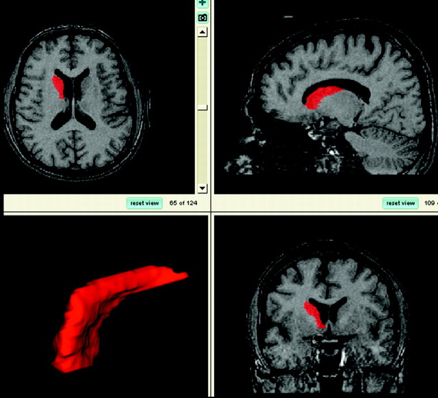
Materials and methods: Diagnoses were based on accepted clinical criteria. Manual volume measurement of the head and body of the caudate, excluding the tail, was conducted on T1-weighted brain MR imaging scans, using a published protocol, by a single analyst blinded to the diagnosis.
Results: Paired t tests (P < .05) showed that the right caudate nucleus volume was significantly larger than the left in controls and PNFA. No hemispheric asymmetry was found in AD, FTD, and SD. Across the groups, there was a positive partial correlation between the left caudate nucleus volume and Mini-Mental State Examination (MMSE) scores (r = 0.393, n = 76, P = .001) with higher left caudate volumes associated with higher MMSE scores. Multivariate analysis of covariance was used to assess the statistical significance between the subject groups (AD, FTD, SD, PNFA, and controls) as independent variables and raw right/left caudate volumes at the within-subject level (covariates: age and intracranial volume; P < .05). Control volume was largest, followed by AD (93% of control volume), SD (92%), PNFA (79%), and FTD (75%).
Conclusions: Volume of the head and body of the caudate nucleus differs in subtypes of FTLD, due to differential frontostriatal dysfunction in subtypes being reflected in structural change in the caudate, and is correlated with cognition.
Figures
Similar articles
-
Putaminal volume in frontotemporal lobar degeneration and Alzheimer disease: differential volumes in dementia subtypes and controls.AJNR Am J Neuroradiol. 2009 Sep;30(8):1552-60. doi: 10.3174/ajnr.A1640. Epub 2009 Jun 4. AJNR Am J Neuroradiol. 2009. PMID: 19497964 Free PMC article.
-
Shape analysis of the neostriatum in subtypes of frontotemporal lobar degeneration: neuroanatomically significant regional morphologic change.Psychiatry Res. 2011 Feb 28;191(2):98-111. doi: 10.1016/j.pscychresns.2010.09.014. Epub 2011 Jan 15. Psychiatry Res. 2011. PMID: 21237621
-
Cortical morphometric subclassification of frontotemporal lobar degeneration.AJNR Am J Neuroradiol. 2009 Jun;30(6):1233-9. doi: 10.3174/ajnr.A1545. Epub 2009 Apr 3. AJNR Am J Neuroradiol. 2009. PMID: 19346314 Free PMC article.
-
Frontotemporal lobar degeneration: clinical and pathological relationships.Acta Neuropathol. 2007 Jul;114(1):31-8. doi: 10.1007/s00401-007-0236-3. Epub 2007 Jun 14. Acta Neuropathol. 2007. PMID: 17569065 Review.
-
[Brain functional imaging of frontotemporal lobar degeneration].Brain Nerve. 2009 Nov;61(11):1275-84. Brain Nerve. 2009. PMID: 19938684 Review. Japanese.
Cited by
-
Regional infant brain development: an MRI-based morphometric analysis in 3 to 13 month olds.Cereb Cortex. 2013 Sep;23(9):2100-17. doi: 10.1093/cercor/bhs197. Epub 2012 Jul 6. Cereb Cortex. 2013. PMID: 22772652 Free PMC article.
-
Reduced caudate nuclei volumes in patients with congenital central hypoventilation syndrome.Neuroscience. 2009 Nov 10;163(4):1373-9. doi: 10.1016/j.neuroscience.2009.07.038. Epub 2009 Jul 24. Neuroscience. 2009. PMID: 19632307 Free PMC article.
-
Shape abnormalities of the caudate nucleus correlate with poorer gait and balance: results from a subset of the LADIS study.Am J Geriatr Psychiatry. 2015 Jan;23(1):59-71.e1. doi: 10.1016/j.jagp.2013.04.011. Epub 2013 Aug 1. Am J Geriatr Psychiatry. 2015. PMID: 23916546 Free PMC article.
-
Looking beneath the surface: the importance of subcortical structures in frontotemporal dementia.Brain Commun. 2021 Jul 16;3(3):fcab158. doi: 10.1093/braincomms/fcab158. eCollection 2021. Brain Commun. 2021. PMID: 34458729 Free PMC article. Review.
-
Fronto-Striatal Atrophy in Behavioral Variant Frontotemporal Dementia and Alzheimer's Disease.Front Neurol. 2015 Jul 1;6:147. doi: 10.3389/fneur.2015.00147. eCollection 2015. Front Neurol. 2015. PMID: 26191038 Free PMC article.
References
-
- Schroeter ML, Razcka K, Neumann J, et al. Towards a nosology for frontotemporal lobar degenerations: a meta-analysis involving 267 subjects. Neuroimage 2007;36:497–510 - PubMed
-
- Postuma RB, Dagher A. Basal ganglia functional connectivity based on a meta-analysis of 126 positron emission tomography and functional magnetic resonance imaging publications. Cereb Cortex 2007;16:1508–21 - PubMed
-
- von Braunmühl A, Picksche K. In: Bumke O, ed. Handbuch der Geisteskrankheiten. Vol. 11. Part VII. Berlin, Germany: Springer-Verlag;1930. :673–715
-
- von Bagh K. Klinische und pathologisch-anatomische Studien an 30 Fällen systematischer umschriebener Atrophie der Grosshirnrinde (Pickscher Krankheit): Annales Academiae Scientiarum Fennicae—Series A. V. Medica Anthropologica Helsinki. Helsinki, Finland: Suomalaisen Tiedeakatemia;1946
Publication types
MeSH terms
LinkOut - more resources
Full Text Sources
Medical