Review
. 2009 Apr;11(4):765-76.
doi: 10.1089/ars.2008.2221.

TRPing on the lung endothelium: calcium channels that regulate barrier function

Affiliations
Review

TRPing on the lung endothelium: calcium channels that regulate barrier function

Donna L Cioffi et al. Antioxid Redox Signal. 2009 Apr.

Abstract

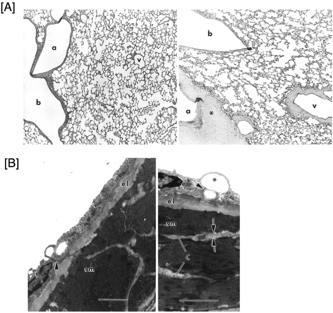
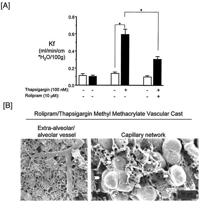
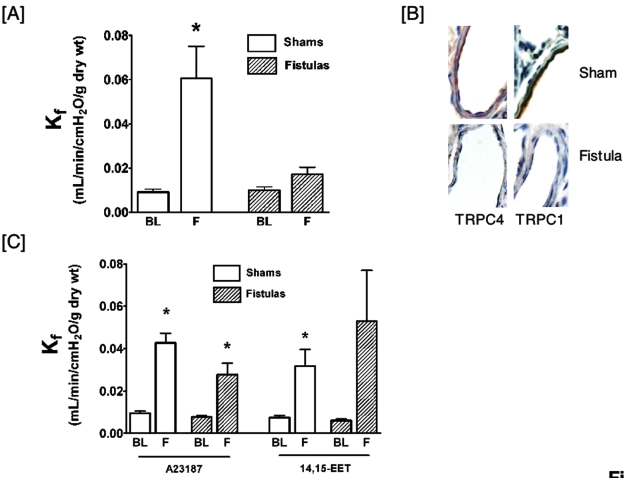
Rises in cytosolic calcium are sufficient to initiate the retraction of endothelial cell borders and to increase macromolecular permeability. Although endothelial cell biologists have recognized the importance of shifts in cytosolic calcium for several decades, only recently have we gained a rudimentary understanding of the membrane calcium channels that change cell shape. Members of the transient receptor potential family (TRP) are chief among the molecular candidates for permeability-coupled calcium channels. Activation of calcium entry through store-operated calcium entry channels, most notably TRPC1 and TRPC4, increases lung endothelial cell permeability, as does activation of calcium entry through the TRPV4 channel. However, TRPC1 and TRPC4 channels appear to influence the lung extraalveolar endothelial barrier most prominently, whereas TRPV4 channels appear to influence the lung capillary endothelial barrier most prominently. Thus, phenotypic heterogeneity in ion channel expression and function exists within the lung endothelium, along the arterial-capillary-venous axis, and is coupled to discrete control of endothelial barrier function.


Figures

FIG. 1.
FIG. 1.
Activation of SOC entry increases endothelial cell permeability. (A) Pulmonary artery endothelial cells grown to confluence were loaded with Fura2/AM, and cytosolic calcium measured using the recalcification protocol. Thapsigargin (TG; 1 μM) applied to cells incubated in nominal extracellular calcium (100 nM) induced a transient increase in cytosolic calcium, due to calcium release from the ER. Replenishing extracellular calcium (Ca2+) to 2 mM resulted in the rapid influx of calcium through open SOC entry channels. For experimental details, see ref. . (B) Pulmonary artery endothelial cells were grown to confluence on transwell plates, and dextran permeability was measured across the monolayer over a 75-min time period. Application of thapsigargin to cells in the low extracellular calcium (100 nM) did not increase macromolecular permeability. However, replenishing extracellular calcium, to allow calcium entry through SOC entry channels, abruptly increased permeability. For experimental details, see (50).
FIG. 2.
FIG. 2.
A spectrin–protein 4.1 interaction is necessary for ISOC activation. (A) Schematic representing the actin and protein 4.1 binding sites on nonerythroid β-spectrin. The SG48 antibody targets a distal region within the protein 4.1 binding domain, and disrupts the spectrin–protein 4.1 interaction. (B) Microinjection of SG48 into pulmonary artery endothelial cells decreases the global cytosolic calcium response to thapsigargin (1 μM). For experimental details, see ref. . (C) Microinjection of SG48 into single pulmonary artery endothelial cells abolishes thapsigargin (1 μM) activation of ISOC. For experimental details, see ref. . (For interpretation of the references to color in this figure legend, the reader is referred to the web version of this article at www.liebertonline.com/ars).
FIG. 3.
FIG. 3.
Schematic representation of the TRP protein family tree. TRP proteins were first identified in Drosophila melanogaster. The canonical (TRPC) mammalian proteins possess the highest homology to Drosophila TRPs. Greater divergence is seen in the melastatin (TRPM) and vanilloid (TRPV) subfamilies. Adapted from ref. . Reprinted, with permission, from the Annual Review of Chemistry, Volume 76 ©2007 by Annual Reviews www.annualreviews.org (For interpretation of the references to color in this figure legend, the reader is referred to the web version of this article at www.liebertonline.com/ars).
FIG. 4.
FIG. 4.
TRPC4 and protein 4.1 interact in endothelial cells. (A) Conserved protein 4.1 binding domains (blue) were identified on the TRPC3 and TRPC4 carboxy-tails, downstream of the signature sequence that is conserved in all TRPC proteins (green) and a proline-rich region (red). (B) TRPC4 and protein 4.1 show similar salt-sensitivity in detergent extractions. Detergent extraction and immuno-blotting was performed over a range of salt (potassium iodide, KI) concentrations. Whereas both TRPC4 and protein 4.1 are resolved in the pellet fraction in the absence of salt, 100 mM KI is sufficient to dissociate TRPC4 and protein 4.1 into the supernatant fraction. For experimental details, see ref. . (C) TRPC4 and protein 4.1 co-immunoprecipitate in the salt-dissociated supernatant. For experimental details, see ref. . (For interpretation of the references to color in this figure legend, the reader is referred to the web version of this article at www.liebertonline.com/ars).
FIG. 5.
FIG. 5.
Thapsigargin induces leak sites in extra-alveolar vessels. (A) Thapsigargin (100 nM) infusion into the circulation of isolated perfused lungs results in increased lung permeability, with abundant fluid accumulation in cuffs around arteries and veins. Left panel is a vehicle control lung section and the right panel is a thapsigargin-treated lung section. a, artery; b, bronchiole; v, vein. For experimental details, see (12). (B) Morphological assessment revealed that thapsigargin induces leak sites in extra-alveolar vessels. Transmission electron micrographs show subendothelial fluid accumulation with thickening of the subendothelial architecture and distention of collagen bundles between adjacent smooth muscle cells. C, collagen; el, elastic lamina; sm, smooth muscle; arrowhead denotes a partially preserved junction. *bleb, indicative of endothelial leak. For experimental details, see ref. .
FIG. 6.
FIG. 6.
Thapsigargin increases segment-specific permeability. (A) Thapsigargin application to isolated perfused rat lungs increases permeability, as determined by the filtration coefficient (Kf). The type 4 phosphodiesterase inhibitor, rolipram, increases endothelial cell cAMP and reduces whole lung permeability. (B) Whereas thapsigargin induces leak sites in extra-alveolar vessel segments (see Figure 5), rolipram pretreatment prevents thapsigargin from inducing extra-alveolar leak. Methyl methacrylate casting material was perfused through rat lungs after thapsigargin and rolipram treatment. After hardening, the cast depicts leak sites that can be resolved by scanning electron microscopy. A precapillary segment and a capillary plexus are shown on the left panel. Whereas leak sites are absent in the precapillary segment, leaks sites are resolved as bulges of the casting material in the capillary segment. Bulges of the casting material (arrowheads) are shown at higher magnification on the right panel. For experimental details, see ref. .
FIG. 7.
FIG. 7.
Lungs from animals with chronic heart failure do not respond to thapsigargin with an increase in permeability, whereas they do respond to 14,15-EET with an increase in permeability. (A) Thapsigargin increases permeability in isolated lungs from sham-operated animals, but does not increase permeability in isolated lungs from animals chronically adapted to an aortocaval fistula. BL, baseline Kf; F, final Kf after thapsigargin infusion. For experimental details, see (2). (B) TRPC1 and TRPC4 are downregulated in endothelium from the lungs of animals with chronic heart failure. Immunostains from sham-treated animals and animals receiving aortocaval shunts show decreased immunoreactivity in the endothelium. (C) Calcium ionophore (A23187) and 14,15-EET increase permeability in both sham-operated and aortaocaval-containing animals. Kf was measured in lungs from control animals and animals with heart failure. In both cases, A23187 and 14,15-EET increased permeability. BL, baseline Kf; and F, final Kf after thapsigargin infusion. For experimental details, see ref. . (For interpretation of the references to color in this figure legend, the reader is referred to the web version of this article at www.liebertonline.com/ars).

Similar articles

Cited by

References

    1. Alvarez D. King J. Weber D. Addison E. Liedtke W. Townsley M. Transient receptor potential vanilloid 4-mediated disruption of the alveolar septal barrier: a novel mechanism of acute lung injury. Circ Res. 2006;99:915–917. - PMC - PubMed
    1. Alvarez DF. King JA. Townsley MI. Resistance to store depletion-induced endothelial injury in rat lung after chronic heart failure. Am J Respir Crit Care Med. 2005;172:1153–1160. - PMC - PubMed
    1. Anderson JP. Morrow JS. The interaction of calmodulin with human erythrocyte spectrin. Inhibition of protein 4.1-stimulated actin binding. J Biol Chem. 1987;262:6365–6372. - PubMed
    1. Antoniotti S. Fiorio Pla A. Barral S. Scalabrino O. Munaron L. Lovisolo D. Interaction between TRPC channel subunits in endothelial cells. J Recept Signal Transduct Res. 2006;26:225–240. - PubMed
    1. Babcock DF. Herrington J. Goodwin PC. Park YB. Hille B. Mitochondrial participation in the intracellular Ca2+ network. J Cell Biol. 1997;136:833–844. - PMC - PubMed

Publication types

MeSH terms

Substances

LinkOut - more resources