Comparative Study
. 2008 Sep 11:8:69.
doi: 10.1186/1472-6750-8-69.

Measuring microRNAs: comparisons of microarray and quantitative PCR measurements, and of different total RNA prep methods

Affiliations
Comparative Study

Measuring microRNAs: comparisons of microarray and quantitative PCR measurements, and of different total RNA prep methods

Robert A Ach et al. BMC Biotechnol. .

Abstract

Background: Determining the expression levels of microRNAs (miRNAs) is of great interest to researchers in many areas of biology, given the significant roles these molecules play in cellular regulation. Two common methods for measuring miRNAs in a total RNA sample are microarrays and quantitative RT-PCR (qPCR). To understand the results of studies that use these two different techniques to measure miRNAs, it is important to understand how well the results of these two analysis methods correlate. Since both methods use total RNA as a starting material, it is also critical to understand how measurement of miRNAs might be affected by the particular method of total RNA preparation used.

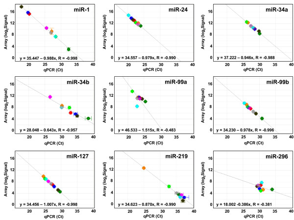
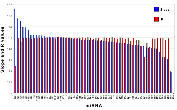
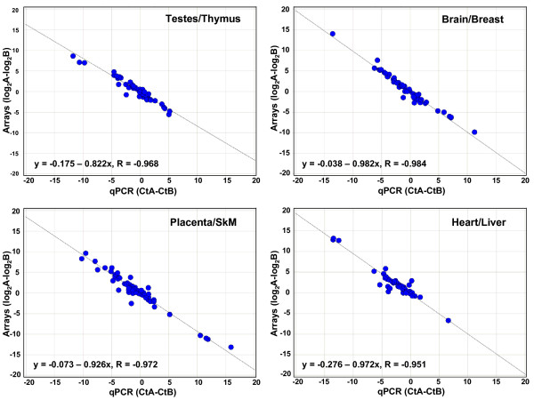
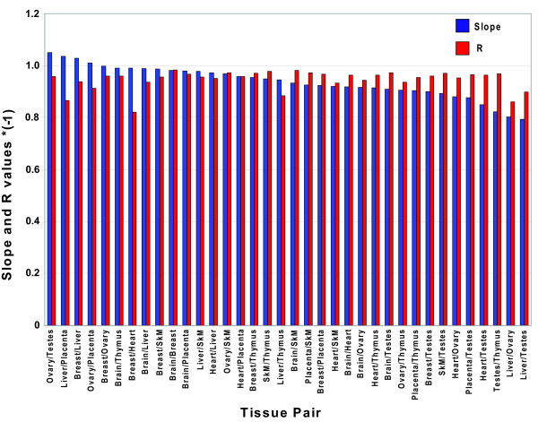
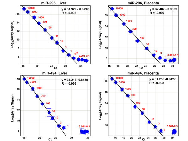
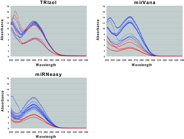
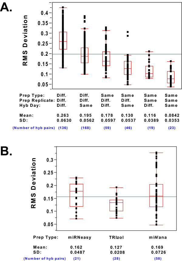
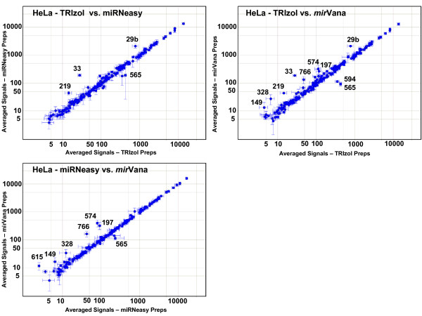
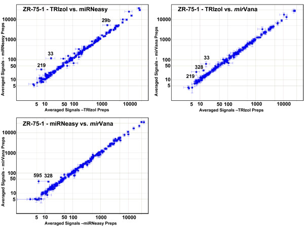
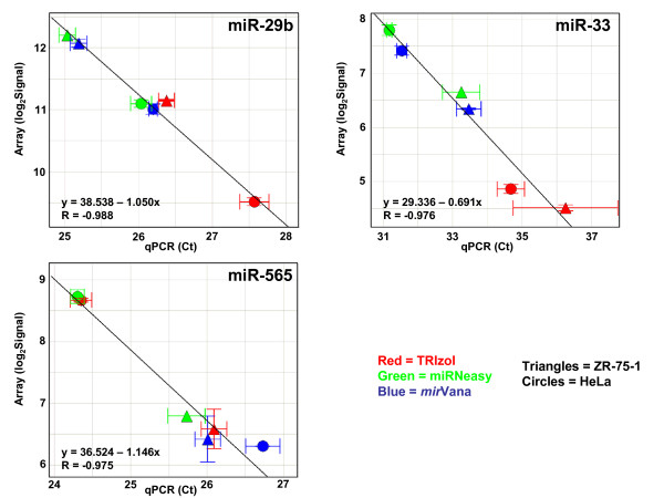
Results: We measured the expression of 470 human miRNAs in nine human tissues using Agilent microarrays, and compared these results to qPCR profiles of 61 miRNAs in the same tissues. Most expressed miRNAs (53/60) correlated well (R > 0.9) between the two methods. Using spiked-in synthetic miRNAs, we further examined the two miRNAs with the lowest correlations, and found the differences cannot be attributed to differential sensitivity of the two methods. We also tested three widely-used total RNA sample prep methods using miRNA microarrays. We found that while almost all miRNA levels correspond between the three methods, there were a few miRNAs whose levels consistently differed between the different prep techniques when measured by microarray analysis. These differences were corroborated by qPCR measurements.

Conclusion: The correlations between Agilent miRNA microarray results and qPCR results are generally excellent, as are the correlations between different total RNA prep methods. However, there are a few miRNAs whose levels do not correlate between the microarray and qPCR measurements, or between different sample prep methods. Researchers should therefore take care when comparing results obtained using different analysis or sample preparation methods.


Figures

Figure 1
Figure 1
Comparison of qPCR and microarray miRNA profiling for individual miRNAs in nine human tissues. Scatter plots are shown for 9 of the 61 miRNAs assayed, with qPCR results (cycle threshold (Ct) values) on the x axes and microarray results (log2 of the total gene signal) on the y axes. Each data point represents one tissue. All plots are drawn to the same scale. The equations and R values on each plot are for the orthogonally-fitted line. Spot colors indicate the tissue: red = breast, pink = testes, dark blue = heart, light blue = placenta, dark green = liver, light green = ovary, orange = brain, brown = skeletal muscle, and grey = thymus. Tissues where qPCR results were flagged as "undetermined" by ABI software, or where log2 of the total gene signal on arrays was < 1, were not plotted. Error bars indicate standard deviation (SD) of Ct values for qPCR results and (SD/Mean)*log2e of the signals for the array results. Scatter plots for the remaining 51 miRNAs (one miRNA gave no signals with either qPCR or arrays) are in Additional Files 2, 3, 4, 5, 6, 7.
Figure 2
Figure 2
Slope and R values for all 60 miRNA scatter plots. The slopes and R values for the orthogonal fit lines for all 60 of the miRNAs plotted in Figure 1 and Additional Files 2, 3, 4, 5, 6, 7 are ordered by the slope values. Slopes are shown in blue and R values are in red.
Figure 3
Figure 3
Comparison of qPCR and microarray miRNA measurements for 60 miRNAs in four tissue pairs. Scatter plots are shown for miRNA expression ratios in four different tissue pairs, as determined by qPCR (x axis) and microarrays (y axis), where each data point represents one miRNA. The qPCR values are the difference between the Ct values from the two tissues, and the microarray values are the difference between the log2(total gene signals) from the two tissues. The equations and R values on each plot are for the orthogonally-fitted line. Scatter plots for the remaining 32 tissue pairs are shown in Additional Files 8, 9, 10, 11, 12, 13, 14, 15. SkM = Skeletal Muscle.
Figure 4
Figure 4
Slope and R values for all 36 tissue pair plots. The slopes and R values for the orthogonal fit lines for all 36 possible tissue pairs plotted in Figure 3 and Additional Files 8, 9, 10, 11, 12, 13, 14, 15 are ordered by the slope values. Slopes are shown in blue and R values are in red.
Figure 5
Figure 5
Comparison of qPCR and microarray measurements for miR-296 and miR-494 titrations. Scatter plots are shown for titration of synthetic miR-296 into liver (top left panel) and placenta (top right panel) total RNAs, and miR-494 into liver (lower left) and placenta (lower right) total RNAs. Ct values from qPCR are plotted on the x-axis, while log2 of the total gene signal from microarray measurements are plotted on the y-axis. Numbers in red show the number of attomoles of spike-in miRNA per 100 ng total RNA. Equations and R values are for the orthogonal line fit of the linear regions of each titration. Error bars indicate standard deviation (SD) of Ct values for qPCR results and (SD/Mean)*log2e for the array results.
Figure 6
Figure 6
Absorption Spectra of HeLa and ZR-75-1 Sample Preps. The absorption spectra for the total RNA sample preps (listed in Table 1) are shown. Blue traces are HeLa cell preps and red traces are ZR-75-1 cell preps. Spectra are from 220 to 350 nm.
Figure 7
Figure 7
Box plots of RMS deviations for microarray hybridization pairs. (a) RMS deviations of all possible hybridization pairs of the same cell type. Box plots show the distribution of RMS deviations calculated from all possible pairs of hybridizations done with the same cell type, and sorted by prep type, prep replicate, and hybridization day. Plots show the combined results from HeLa and ZR-75-1 cells. Each replicate pair RMS deviation is indicated by a black dot. The ends of the box represent the 25th and 75th quartiles, while the line through the center of the box represents the median. The whiskers from each box extend to the outermost data point included in the range from the upper quartile plus 1.5*(interquartile range) to the lower quartile minus 1.5*(interquartile range). The line across the entire plot indicates the mean of all the values. Values of the mean and standard deviations for each set of values are shown at the bottom of the plot. (b) RMS deviations of all prep replicate hybridization pairs, sorted by prep type. Box plots of the RMS deviations for all the pair-wise comparisons of hybridizations using replicates of the same prep type, and sorted by prep type. Plots show the combined results from HeLa and ZR-75-1 cells. Box plot details are as in Figure 7a.
Figure 8
Figure 8
Pair-wise comparisons of averaged profiles of the three different prep types: HeLa cells. Total gene signals for each miRNA for all hybridizations of the same RNA prep (hybridization replicates) were averaged, and then these averaged individual prep profiles for all the preps of the same prep type were averaged together to get a mean profile for each of the three prep types. Scatter plots show these averaged profiles from one prep type plotted against another for HeLa cells. Error bars indicate one standard deviation. Numbers indicate the identity of all miRNAs whose signal strengths are at least two-fold higher in one prep type than another, after normalization.
Figure 9
Figure 9
Pair-wise comparisons of averaged profiles of the three different prep types: ZR-75-1 preps. Scatter plots as in Figure 8, but for ZR-75-1 cell preps.
Figure 10
Figure 10
Comparison of qPCR and microarray miRNA results for three miRNAs differentially measured between different prep types. Individual TRIzol, miRNeasy, and mirVana preps were assayed with qPCR for three miRNAs found in microarray studies to be at higher levels in one prep type than another. Scatter plots show qPCR results (cycle threshold (Ct) values) on the x axes and microarray results (log2 of the total gene signal) on the y axes. Each data point represents one individual prep from one cell type. Circles indicate HeLa preps and triangles represent ZR-75-1 preps. TRIzol preps are in red, miRNeasy preps are in green, and mirVana preps are in blue. The equations and R values on each plot are for the line of best fit. Error bars indicate standard deviation (SD) of Ct values for qPCR results and (SD/Mean)*log2e for the array results. Note that the axes are not on the same scale in the three different plots.

Similar articles

Cited by

References

    1. Nilsen TW. Mechanisms of microRNA-mediated gene regulation in animal cells. Trends Genet. 2007;23:243–249. doi: 10.1016/j.tig.2007.02.011. - DOI - PubMed
    1. Pillai RS, Bhattacharyya SN, Filipowicz W. Repression of protein synthesis by miRNAs: How many mechanisms? Trends Cell Biol. 2007;17:118–126. doi: 10.1016/j.tcb.2006.12.007. - DOI - PubMed
    1. Eulalio A, Huntzinger E, Izaurralde E. Getting to the root of miRNA-mediated gene silencing. Cell. 2007;132:9–14. doi: 10.1016/j.cell.2007.12.024. - DOI - PubMed
    1. Vasudevan S, Tong Y, Steitz J. Switching from repression to activation: MicroRNAs can up-regulate translation. Science. 2008;318:1931–1934. doi: 10.1126/science.1149460. - DOI - PubMed
    1. Leung AKL, Sharp PA. microRNAs: A safeguard against turmoil? Cell. 2007;130:581–585. doi: 10.1016/j.cell.2007.08.010. - DOI - PubMed

Publication types

MeSH terms