Review
. 2008 Oct;29(5):309-28.
doi: 10.1016/j.mam.2008.08.002. Epub 2008 Aug 22.

Meprins, membrane-bound and secreted astacin metalloproteinases

Affiliations
Review

Meprins, membrane-bound and secreted astacin metalloproteinases

Erwin E Sterchi et al. Mol Aspects Med. 2008 Oct.

Abstract

The astacins are a subfamily of the metzincin superfamily of metalloproteinases. The first to be characterized was the crayfish enzyme astacin. To date more than 200 members of this family have been identified in species ranging from bacteria to humans. Astacins are involved in developmental morphogenesis, matrix assembly, tissue differentiation and digestion. Family members include the procollagen C-proteinase (BMP1, bone morphogenetic protein 1), tolloid and mammalian tolloid-like, HMP (Hydra vulgaris metalloproteinase), sea urchin BP10 (blastula protein) and SPAN (Strongylocentrotus purpuratus astacin), the 'hatching' subfamily comprising alveolin, ovastacin, LCE, HCE ('low' and 'high' choriolytic enzymes), nephrosin (from carp head kidney), UVS.2 from frog, and the meprins. In the human and mouse genomes, there are six astacin family genes (two meprins, three BMP1/tolloid-like, one ovastacin), but in Caenorhabditis elegans there are 40. Meprins are the only astacin proteinases that function on the membrane and extracellularly by virtue of the fact that they can be membrane-bound or secreted. They are unique in their domain structure and covalent subunit dimerization, oligomerization propensities, and expression patterns. They are normally highly regulated at the transcriptional and post-translational levels, localize to specific membranes or extracellular spaces, and can hydrolyse biologically active peptides, cytokines, extracellular matrix (ECM) proteins and cell-surface proteins. The in vivo substrates of meprins are unknown, but the abundant expression of these proteinases in the epithelial cells of the intestine, kidney and skin provide clues to their functions.


Figures

Figure 1
Figure 1. Proteases, metzincins, astacins, meprins
Schematic overview of proteolytic enzymes with special emphasis on metzincin metalloproteinases. The superscripts indicate the numbers of human and mouse protease genes (http://merops.sanger.ac.uk). Enzymes are indicated with UniProtKB/Swiss-Prot code. Ser, serine proteases (trypsin P07478; thrombin P00734; furin P09958, matriptase Q9Y5Y6); Cys, cysteine proteinases (cathepsins B P07711, L P07858, K P43235, caspase 8 Q14790, calpain P07384); Thr, threonine proteinases (human proteasome β4 P28070); Asp/Glu, aspartate and glutamate proteinases (pepsin A P00790, HIV proteinase P04586, renin P00797, BACE P56817, γ secretase P49810, SCP-B P15369, i.e. scytalidoglutamic peptidase); Metallo = metalloproteases; Non-Zincins, metalloproteinases with alternate zinc motifs (carboxypeptidase A P00730, DD-carboxypeptidase P00733); Inverzincins, metalloproteinases with the inverted zinc binding motif HXXEH (pitrilysin P05458, insulysin P14735); Zincins, metalloproteinases with the zinc binding consensus sequence HEXXH (single letter code, X = any amino acid residue); Gluzincins, metalloproteinases with the zinc binding motif HEXXH…E (thermolysin P00800; ACE P12821, i.e. angiotensin converting enzyme, Anthrax lethal factor P15917; Tentoxilysin P04958, Bontoxilysin P10844, neprilysin P08473, thimet oligopeptidase P24155); Aspzincins, metalloproteinases withe the zinc binding motif HEXXH.…D (deuterolysin, P46073); Metzincins, metalloproteinases with the zinc binding motif HEXXHXXG/NXXH/D); Leishmanolysins (leishmanolysin, P08148; invadolysin, Q9VH19); Pappalysins (PAPP_A, Q13219); Serralysins (peptidase B P16316, serralysin P23694); ADAMs (A Disintegrin And Metalloproteinase; TACE i.e. ADAM17 P78536, ADAM10 i.e. α secretase Q10741, ADAMTS2 P79331); MMPs (matrix metalloproteinases, MMP1 collagenase P79331, MMP2 gelatinase P08253, MT1-MMP P50281); Astacins (astacin P07584; hook worm AMP1 1 Q9GTJ6, trichina TMP1 Q8T5Z5, Onchocerca onchoastacin Q2YFS7, C. elegans NAS36 Q18206, Hydra HMP1 Q25174, Hydra FARM1; Hydractinia HE2-4 Q2MCX8 Q2MCX7 Q2MCX6, Podecoryne PMP1 O62558, quail CAM1 P42662, carp alveolin Q9IBE7, carp nephrosin O42326, medaka HCE P31580, medaka LCE P31579, frog UVS2 P42664, human ovastacin Q6HA08, fly tolloid P25723, human BMP1 P13497, human Tll1 O43897, human Tll2 Q9Y6L7, sea urchin SPAN P98068, sea urchin BP10 P42674, squid myosinases Q8IU47 Q8IU44 Q8IU46 Q8IU45, Hydra HMP2 Q9XZG0, sea anemone NVE_MAM A7SJ13, fish meprin a1 Q5RHM1, fish meprin a2 Q5RHM2, mouse meprin a P28825, human meprin a Q16819, fish meprin b Q08CC, human meprin b Q16820, mouse meprin b Q61847. Below the different metzincin subfamilies, ribbon plots are depicted of representative catalytic domain structures: leishmanolysin (1lml.pdb), ulilysin (2cki.pdb), serralysin (1srp.pdb), TACE (1bck.pdb), MMP8 (1mnc.pdb), and astacin (1ast.pdb).
Figure 2
Figure 2. Domain structure of human meprins
Meprins consist of an N-terminal signal peptide (S), a prodomain (PRO), a proteinase domain containing the metzincin motif HxxLHxxGxxH, a MAM domain (meprin, A5 protein, receptor protein tyrosine phosphatase µ), a TRAF domain (tumor necrosis factor receptor-associated factor), an epidermal growth factor-like domain (EGF), a transmembrane domain (TM) and a cytosolic tail sequence (C), which is only 6 amino acid residues long in the α subunit and 28 amino acid residues in the β subunit. The α subunit contains an additional inserted sequence (I), which is essential for the constitutive proteolytic processing of this subunit. Three intersubunit S-S bridges are proposed for the human heterodimer; only two intersubunit bridges are possible for the mouse heterodimer because this subunit has no Cys residues in the TRAF domain. Cleavage by trypsin or trypsin-like enzymes results in removal of the prodomain and activation of the meprin subunits (Jiang, Gorbea et al. 1992; Dumermuth, Eldering et al. 1993; Eldering, Grunberg et al. 1997).
Figure 3
Figure 3. Model of the proteinase domain structure of meprins
Based on the structure of astacin (Bode, Gomis-Rüth et al. 1992) (1ast.pdb), the homologous proteinase domain of meprins consists of a five-stranded β-sheet, three α-helices and coil structure in the lower subdomain (Stöcker, Gomis-Rüth et al. 1993). The catalytic zinc (large black sphere) is penta-coordinated by three histidines, the catalytic water (small black sphere) and a tyrosine positioned by the Met-turn, containing the eponymous methionine typical for metzincin proteinases. The structure of the catalytic domain is stabilized by two intradomain disulfide-bridges and several conserved salt bridges.
Figure 4
Figure 4. Model of the meprin β dimer
A model of the dimeric structure is proposed based upon disulfide and cross-linking mapping experiments. The dimmer exhibits D2 symmetry and is arranged such that the subunit interface is formed between the MAM domains, the TRAF domains, and the prosequences. The protease domain interacts with the TRAF domain as well as the EGF domain, placing it near the COOH-terminal region of the protein close to the cell membrane (Ishmael, Shier et al. 2005).
Figure 5
Figure 5. Oligomerization of meprin proteinases
α Electron micrographs of meprin α expressed in human embryonic kidney 293 show disulphide-bridged homodimers which can further associate noncovalently in long, curved chains to form rings and spirals of variable sizes that reach molecular masses in the mega Dalton range. By contrast, meprin β homooligomers form only disulphide-linked dimers that are incapable of further aggregation. α/β When coexpressed, meprin α and β subunits show disulphide-linked heterodimers which mostly associate non-covalently as heterotetramers. Below the electron micrographs the oligomeric arrangements of the individual subunits is schematically depicted (Becker, Kruse et al. 2003; Bertenshaw, Norcum et al. 2003).
Figure 6
Figure 6. Gene organization of meprins
The top line in each panel shows a schematic diagram of the respective gene. Exons are numbered and are represented as follows: black boxes, coding regions; open boxes, non-coding regions. Introns are presented as dashes. pKS clones containing genomic sequences are shown above the line. Arrows indicate the position and direction of primers used during the sequencing project. The bottom line in each panel demonstrates a schematic diagram of meprin α and meprin β protein derived from the exons. The amino acid numbers at the beginning of each domain are also presented. Abbreviations : H, Hin dIII ; E, Eco RI ; P, Pst I. Regions of the protein are indicated as follows: formula image 5’ untranslated region; formula image signal peptide; formula image propeptide; proteinase; formula image MAM (meprin subunit domain/A5 protein/receptor protein tyrosine phosphatase l domain); formula image MATH (meprin- and tumour-necrosis-factor-receptor-associated factors homology domain); formula image intervening domain; formula image I-domain (inserted domain); formula image EGF-like domain; formula image transmembrane domain; formula image cytosolic domain; formula image 3’ untranslated region (Hahn, Illisson et al. 2000).
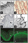
Figure 7
Figure 7. Substrate and Inhibitor specificity of meprins
At the top the strikingly different subsite specificities of mouse meprin α and β are depicted as a WEBLOGO (http://weblogo.berkeley.edu/). The size of the letters reflects the relative abundance of the respective single letter coded amino acid residues in substrate proteins at the P4 through P4’ position (Schechter and Berger 1967). The substrates analyzed are those listed in the paper by (Bertenshaw, Turk et al. 2001). Listed below are substrates and inhibitors of meprin subunits (Bertenshaw, Turk et al. 2001; Kruse, Becker et al. 2004; Herzog, Kaushal et al. 2005; Hirano, Ma et al. 2005).
Figure 8
Figure 8. Tissue expression of meprins
A Immuno-electronmicrograph of proximal tubular cells from ICR mice showing brush border membrane localization of meprin α (Craig, Reckelhoff et al. 1987). B Immunohistochemical staining for meprin β in the brush border membrane of small intestinal epithelial cells (Lottaz, Hahn et al. 1999). C In situ hybridization for meprin mRNA α in small intestinal mucosa from a patient with active coeliac disease. The presence of meprin α shifts from the epithelial cells to leukocytes in the lamina propria (Lottaz, Buri et al. 2007). D In situ hybridization for meprin α mRNA in normal human colon (Lottaz, Hahn et al. 1999). E and F show immunofluorescence staining of human skin for meprin α and β, respectively. The continuous line indicates the outer most border of the stratum corneum, the dashed line indicates the basal membrane of the epidermis. Panel E: Perinuclear fluorescence signal (white arrow head) for meprin α in cells of the stratum basale. Panel F: Signal for meprin β (white arrow head) in membranes of cells of the stratum granulosum beneath the stratum corneum (black arrow head) (Becker-Pauly, Höwel et al. 2007).

Similar articles

Cited by

References

    1. Ambort D, Stalder D, Lottaz D, Hguenin M, Oneda B, Heller M, Sterchi EE. A novel 2D-based approach to the discovery of candidate substrates for the metalloendopeptidase meprin. FEBS J. 2008 - PubMed
    1. Aricescu AR, Hon WC, Siebold C, Lu W, van der Merwe PA, Jones EY. Molecular analysis of receptor protein tyrosine phosphatase mu-mediated cell adhesion. Embo J. 2006;25(4):701–712. - PMC - PubMed
    1. Bankus JM, Bond JS. Expression and distribution of meprin protease subunits in mouse intestine. Arch. Biochem. Biophys. 1996;331(1):87–94. - PubMed
    1. Barnes K, Ingram J, Kenny A. Proteins of the kidney microvillar membrane. Structural and immunochemical properties of rat endopeptidase-2 and its immunohistochemical localization in tissues of rat and mouse. Biochem. J. 1989;264(2):335–346. - PMC - PubMed
    1. Barrett AJ, Rawlings ND, O'Brien EA. The MEROPS database as a protease information system. J. Struct. Biol. 2001;134(2–3):95–102. - PubMed

Publication types