

Spinal cryptococcoma in an immunocompetent cat
- PMID: 18783789
- PMCID: PMC7094615
- DOI: 10.1016/j.jcpa.2008.06.007
Spinal cryptococcoma in an immunocompetent cat
Abstract
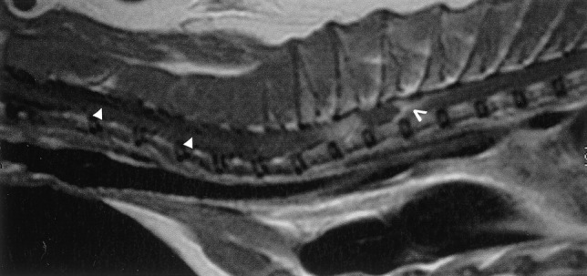
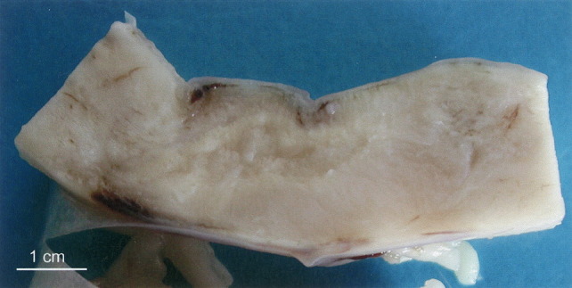
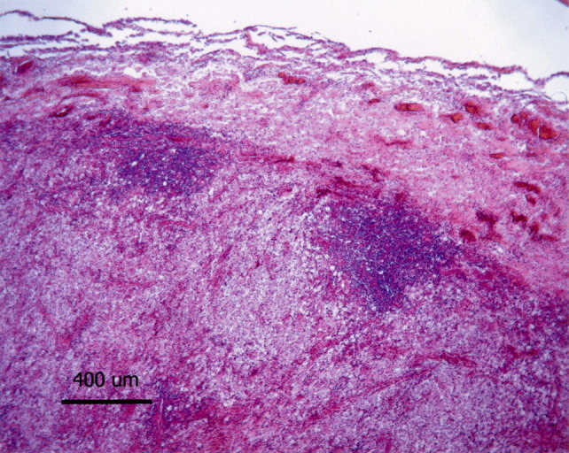
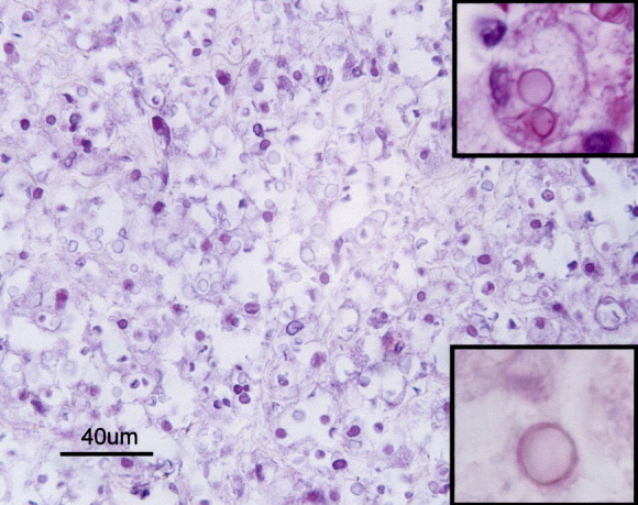
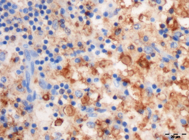
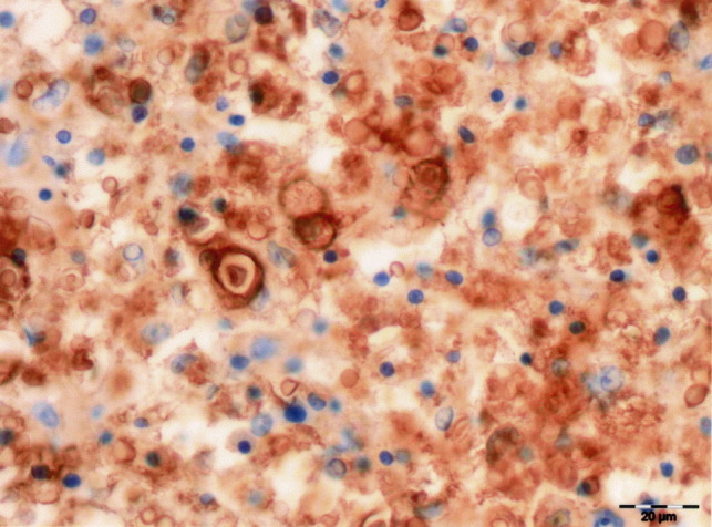
This report describes an unusual case of primary cryptococcoma in the proximal thoracic spinal cord of an 11-year-old immunocompetent cat from a farm on which there were large numbers of pigeons. This animal was referred for examination with progressive paralysis and shown to be free from feline immunodeficiency virus, feline leukaemia virus, feline coronavirus and Toxoplasma gondii. It died 2 months later. At necropsy, the only lesion detected was a malacic area, 4cm in length, in the spinal cord. Histopathological examination of the spinal cord revealed severe granulomatous inflammation associated with large numbers of encapsulated yeast cells. In addition to the granulomatous host response, necrosis, digestion chambers, Gitter cells, spheroids and lymphocytic perivascular cuffs were features of the malacic areas. Immunohistochemistry confirmed the presence of Cryptococcus neoformans var. grubii yeast cells.
Figures
References
-
- Baker R.D. The primary pulmonary lymph node complex of crytptococcosis. American Journal of Clinical Pathology. 1976;65:83–92. - PubMed
-
- Bicanic T., Harrison T.S. Cryptococcal meningitis. British Medical Bulletin. 2004;72:99–118. - PubMed
-
- Caswell J.L., Williams K.J. Cryptococcosis. In: Maxie M.G., editor. vol. 2. Elsevier Saunders; Philadelphia: 2007. pp. 642–644. (Pathology of Domestic Animals).
-
- Chen S., Sorrell T., Nimmo G., Speed B., Currie B., Ellis D., Marriott D., Pfeiffer T., Parr D., Byth K. Epidemiology and host- and variety-dependent characteristics of infection due to Cryptococcus neoformans in Australia and New Zealand. Clinical Infectious Diseases. 2000;31:499–508. - PubMed
Publication types
MeSH terms
LinkOut - more resources
Full Text Sources
Medical
Miscellaneous
