

Elucidating the mechanisms and loci of neuronal excitation by transcranial magnetic stimulation using a finite element model of a cortical sulcus
- PMID: 18783986
- PMCID: PMC2693370
- DOI: 10.1016/j.clinph.2008.07.248
Elucidating the mechanisms and loci of neuronal excitation by transcranial magnetic stimulation using a finite element model of a cortical sulcus
Abstract
Objective: This work aims to elucidate by what physical mechanisms and where stimulation occurs in the brain during transcranial magnetic stimulation (TMS), taking into account cortical geometry and tissue heterogeneity.
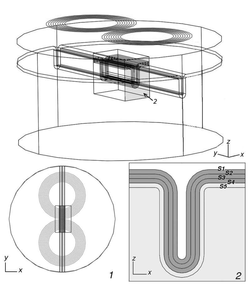
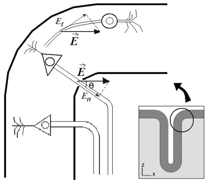
Methods: An idealized computer model of TMS was developed, comprising a stimulation coil, a cortical sulcus, and surrounding tissues. The distribution of the induced electric field was computed, and estimates of the relevant parameters were generated to predict the locus and type of neurons stimulated during TMS, assuming three different stimulation mechanisms.
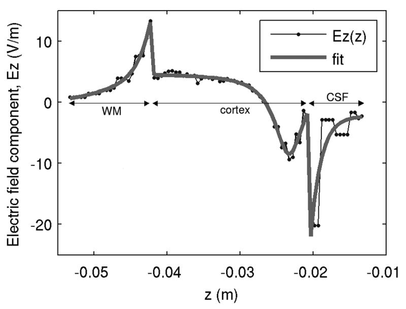
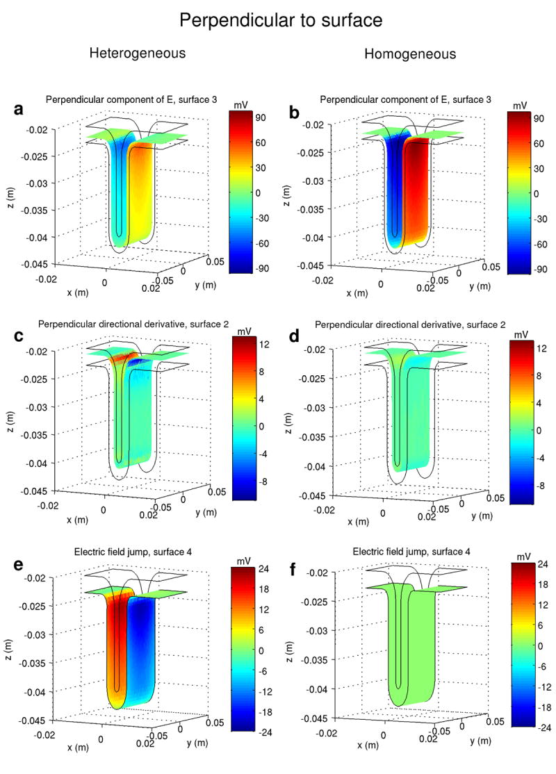
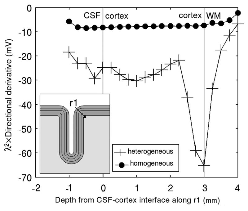
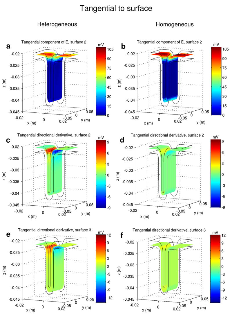
Results: Tissue heterogeneity strongly affects the spatial distribution of the induced electric field and hence which stimulation mechanism is dominant and where it acts. Stimulation of neurons may occur in the gyrus, in the lip of the gyrus, and in the walls of the sulcus. The stimulated cells can be either pyramidal cells having medium to large caliber axons, or intracortical fibers of medium caliber.
Conclusions: The results highlight the influence of cortical folding on the action of magnetic and electric fields on cortical tissue.
Significance: Tissue geometry and heterogeneity in electrical conductivity both must be taken into account to predict accurately stimulation loci and mechanism in TMS.
Figures
Similar articles
-
The activation function of TMS on a finite element model of a cortical sulcus.Annu Int Conf IEEE Eng Med Biol Soc. 2007;2007:6657-60. doi: 10.1109/IEMBS.2007.4353886. Annu Int Conf IEEE Eng Med Biol Soc. 2007. PMID: 18003552
-
Determining which mechanisms lead to activation in the motor cortex: a modeling study of transcranial magnetic stimulation using realistic stimulus waveforms and sulcal geometry.Clin Neurophysiol. 2011 Apr;122(4):748-58. doi: 10.1016/j.clinph.2010.09.022. Epub 2010 Oct 28. Clin Neurophysiol. 2011. PMID: 21035390 Free PMC article.
-
Computational and experimental analysis of TMS-induced electric field vectors critical to neuronal activation.J Neural Eng. 2015 Aug;12(4):046014. doi: 10.1088/1741-2560/12/4/046014. Epub 2015 Jun 8. J Neural Eng. 2015. PMID: 26052136
-
Transcranial magnetic stimulation of the brain: What is stimulated? - A consensus and critical position paper.Clin Neurophysiol. 2022 Aug;140:59-97. doi: 10.1016/j.clinph.2022.04.022. Epub 2022 May 18. Clin Neurophysiol. 2022. PMID: 35738037 Free PMC article. Review.
-
Basic mechanisms of TMS.J Clin Neurophysiol. 2002 Aug;19(4):322-43. doi: 10.1097/00004691-200208000-00006. J Clin Neurophysiol. 2002. PMID: 12436088 Review.
Cited by
-
Localization of Sensorimotor Cortex Using Navigated Transcranial Magnetic Stimulation and Magnetoencephalography.Brain Topogr. 2019 Sep;32(5):873-881. doi: 10.1007/s10548-019-00716-w. Epub 2019 May 15. Brain Topogr. 2019. PMID: 31093863 Free PMC article.
-
Targeting of white matter tracts with transcranial magnetic stimulation.Brain Stimul. 2014 Jan-Feb;7(1):80-4. doi: 10.1016/j.brs.2013.10.001. Epub 2013 Oct 16. Brain Stimul. 2014. PMID: 24220599 Free PMC article.
-
The development and modelling of devices and paradigms for transcranial magnetic stimulation.Int Rev Psychiatry. 2017 Apr;29(2):115-145. doi: 10.1080/09540261.2017.1305949. Epub 2017 Apr 26. Int Rev Psychiatry. 2017. PMID: 28443696 Free PMC article. Review.
-
Rehabilitating the addicted brain with transcranial magnetic stimulation.Nat Rev Neurosci. 2017 Nov;18(11):685-693. doi: 10.1038/nrn.2017.113. Epub 2017 Sep 29. Nat Rev Neurosci. 2017. PMID: 28951609 Review.
-
Adding low-field magnetic stimulation to noninvasive electromagnetic neuromodulatory therapies.Biol Psychiatry. 2014 Aug 1;76(3):170-1. doi: 10.1016/j.biopsych.2014.05.017. Biol Psychiatry. 2014. PMID: 25012043 Free PMC article. No abstract available.
References
-
- Amassian VE, Eberle L, Maccabee PJ, Cracco RQ. Modelling magnetic coil excitation of human cerebral cortex with a peripheral nerve immersed in a brain-shaped volume conductor: the significance of fiber bending in excitation. Electroencephalogr Clin Neurophysiol. 1992;85:291–301. - PubMed
-
- Barker AT, Garnham CW, Freeston IL. Magnetic nerve stimulation: the effect of waveform on efficiency, determination of neural membrane time constants and the measurement of stimulator output. Electroencephalogr Clin Neurophysiol Suppl. 1991;43:227–237. - PubMed
-
- Barker AT, Jalinous R, Freeston IL. Non-invasive magnetic stimulation of human motor cortex. Lancet. 1985;1:1106–1107. - PubMed
-
- Basser PJ, Roth BJ. Stimulation of a myelinated nerve axon by electromagnetic induction. Med Biol Eng Comput. 1991;29:261–268. - PubMed
-
- Baumann SB, Wozny DR, Kelly SK, Meno FM. The electrical conductivity of human cerebrospinal fluid at body temperature. IEEE Trans Biomed Eng. 1997;44:220–223. - PubMed
Publication types
MeSH terms
Grants and funding
LinkOut - more resources
Full Text Sources
