. 2008 Nov 14;283(46):31417-28.
doi: 10.1074/jbc.M806350200. Epub 2008 Sep 10.

Identification of Mycobacterium tuberculosis clinical isolates with altered phagocytosis by human macrophages due to a truncated lipoarabinomannan

Affiliations

Identification of Mycobacterium tuberculosis clinical isolates with altered phagocytosis by human macrophages due to a truncated lipoarabinomannan

Jordi B Torrelles et al. J Biol Chem. .

Abstract

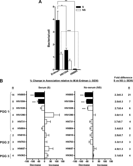
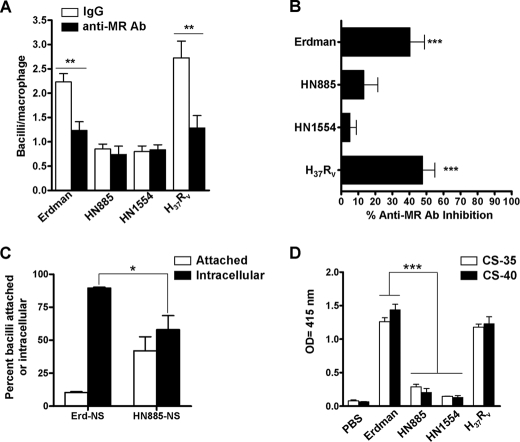
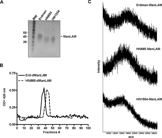
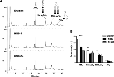
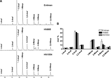
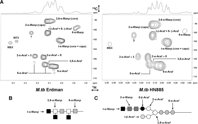
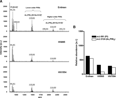
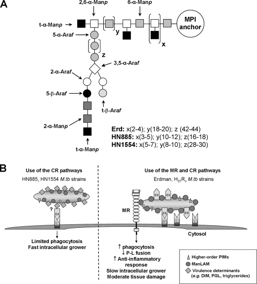
Phenotypically distinct clinical isolates of Mycobacterium tuberculosis are capable of altering the balance that exists between the pathogen and human host and ultimately the outcome of infection. This study has identified two M. tuberculosis strains (i.e. HN885 and HN1554) among a bank of clinical isolates with a striking defect in phagocytosis by primary human macrophages when compared with strain Erdman, a commonly used laboratory strain for studies of pathogenesis. Mass spectrometry in conjunction with NMR studies unequivocally confirmed that both HN885 and HN1554 contain truncated and more branched forms of mannose-capped lipoarabinomannan (ManLAM) with a marked reduction of their linear arabinan (corresponding mainly to the inner Araf-alpha(1-->5)-Araf unit) and mannan (with fewer 6-Manp residues and more substitutions in the linear Manp-alpha(1-->6)-Manp unit) domains. The truncation in the ManLAM molecules produced by strains HN885 and HN1554 led to a significant reduction in their surface availability. In addition, there was a marked reduction of higher order phosphatidyl-myo-inositol mannosides and the presence of dimycocerosates, triglycerides, and phenolic glycolipid in their cell envelope. Less exposed ManLAM and reduced higher order phosphatidyl-myo-inositol mannosides in strains HN885 and HN1554 resulted in their low association with the macrophage mannose receptor. Despite reduced phagocytosis, ingested bacilli replicated at a fast rate following serum opsonization. Our results provide evidence that the clinical spectrum of tuberculosis may be dictated not only by the host but also by the amounts and ratios of surface exposed mycobacterial adherence factors defined by strain genotype.


Figures

FIGURE 1.
FIGURE 1.
Association of M. tuberculosis clinical isolates with human macrophages. MDM monolayers were incubated with Erdman (reference strain) or the indicated M. tuberculosis(M. tb) clinical isolates in the presence (S) or absence (NS) of 2.5% serum for 2 h. Association was measured by phase-contrast and fluorescence microscopy. A, a representative association experiment for HN885 and HN1554 performed by triplicate in the presence or absence of serum. B, data represent percentage association of each clinical isolate relative to Erdman (y axis) for each condition (n is the number of independent experiments with the indicated isolate). *, p < 0.05; **, p < 0.0005; ***, p < 0.0001; Student's t test. The difference in bacterial association between the serum and no serum condition is depicted as summed data on the right.
FIGURE 2.
FIGURE 2.
Macrophage association (A and B), phagocytosis (C), and the exposure of ManLAM on the bacterial surface (D) for strains HN885 and HN1554. A, a representative experiment performed in triplicate in which MDM monolayers were preincubated with 10 μg/ml anti-MR mAb (solid bars) or IgG1 isotype (open bars) before incubation with the M. tuberculosis strains. **, p < 0.005; Student's t test. The results show that HN885 and HN1554 do not use the MR to associate with human macrophages, and the overall MR-dependent association of M. tuberculosis strains with MDMs is represented by percentage inhibition in the presence of anti-MR mAb relative to the subtype control mAb (B). ***, p < 0.0001; one-way analysis of variance Tukey post-test clinical isolates versus M. tuberculosis control strains (Erdman/H37Rv), n = 6. C, percentage of M. tuberculosis Erdman and HN885 attached versus internalized (phagocytosed) by macrophages. MDM monolayers on glass coverslips were incubated with Erdman or HN885 M. tuberculosis in the presence or absence of 2.5% serum for 2 h, fixed, and prepared for TEM analysis. Data represent mean ± S.E. (*, p < 0.05, n = 3 by triplicate) for the percentage of bacteria that were intracellular versus extracellular. D, ManLAM detection on the surface of M. tuberculosis strains. Whole bacterial ELISA using live M. tuberculosis and anti-LAM mAb CS-35 shows a significant reduction in the recognition of surface ManLAM on strains HN885 and HN1554 (***, p < 0.001; one-way analysis of variance, Tukey post-test, clinical isolates versus control strains (Erdman/H37Rv), n = 3 by triplicate).
FIGURE 3.
FIGURE 3.
Size of ManLAMs from M. tuberculosis strains. A, 10–20% gradient Tris/Tricine gel followed by periodic acid-silver staining shows that ManLAMs from strains HN885 and HN1554 exhibit a greater electrophoretic mobility than ManLAM from strain Erdman; the size difference can be approximated by aligning the respective leading edges of the characteristic broad electrophoretic bands. MW, molecular weight. B, sizing column chromatography of deacylated ManLAMs. After deacylation, 1 mg of dManLAM was loaded onto a P-100 sizing column, and 1-ml fractions were collected, dried, and analyzed by carbohydrate content. Erd-dManLAM (solid line) eluted earlier from the column than HN885-dManLAM (interrupted line), indicating that its carbohydrate core is larger. C, negative MALDI mass spectrum of Erdman, HN885, and HN1554 ManLAMs. ManLAM (0.5 μl of a 10 μg/μl solution) was mixed with 0.5 μl of the matrix solution (10 μg/μl) of DHB in ethanol/water (1:1, v/v) and analyzed by MALDI-TOF in the negative mode.
FIGURE 4.
FIGURE 4.
HPAEC profile of endoarabinanase-digested M. tuberculosis ManLAMs. A, representative HPAEC profiles for Erdman, HN885, and HN1554 ManLAMs. For direct comparison, the digestion products were dried and injected directly without further purification. Ara2 and Man2Ara4 are linear oligosaccharides, whereas Ara6 and Man4Ara6 are branched. B, quantification of the peak areas observed by HPAEC analysis. Values represent mean ± S.E. (n = 2) by duplicate. ***, p < 0.0001; Student's t test, clinical isolates versus control strain Erdman.
FIGURE 5.
FIGURE 5.
Linkage analysis of M. tuberculosis ManLAMs as determined by GC/MS. Samples were per-O-methylated, hydrolyzed, reduced, and acetylated, and partially methylated alditol acetates were analyzed by GC/MS as described under “Experimental Procedures.” A, the spectra of various linked Ara and Man derivatives in the ManLAMs from Erdman, HN885, and HN1554 are shown. B, the bar graph shows calculated mol % of specific linked sugars in the purified ManLAMs. Values represent mean ± S.E. (n = 2) by duplicate.
FIGURE 6.
FIGURE 6.
Comparative partial two-dimensional NMR spectra of Erdman and HN885 ManLAMs. A, two-dimensional NMR 1H-13C HSQC spectra were acquired in D2O. Only the expanded anomeric regions are shown.?, unidentified peaks. The intensity of peak volumes was measured, and the data are presented in Table 2. Shown are representations of the sugar linkages described for the mannan (B) and arabinan (C) domains of ManLAM. M. tb, M. tuberculosis.
FIGURE 7.
FIGURE 7.
Total PIM analysis of M. tuberculosis strains by MALDI-TOF. A, the MALDI-TOF mass spectra of the total PIM extract from strains Erdman, HN885, and HN1554. Cell lysates (500 μg normalized by protein content) from each strain were extracted with chloroform/methanol (2:1, v/v) for 12 h, followed by chloroform/methanol (1:2, v/v) for an additional 12 h. Extracts were dried down and precipitated in cold acetone for 12 h. PIMs were mixed with DHB matrix in chloroform/methanol/water (10:10:3, v/v/v) and analyzed by MALDI-TOF in the positive mode. Major differences in lower and higher order PIM content were observed among the M. tuberculosis strains, where strains HN885 and HN1554 contain significantly fewer higher order PIMs than strain Erdman. a.i., arbitrary intensity. B, quantification of PIMs by MALDI-TOF MS. Samples were co-crystallized with DHB matrix on the probe using solvent evaporation, desorbed and ionized by a nitrogen laser pulse (337 nm), and then accelerated under 25 kV with time-delayed extraction before entering the time-of-flight mass spectrometer. The number of laser pulses was received as 3 × 60 or 180 laser pulses for a final MALDI-MS spectrum. Samples with and without the standard (α(1→4)-mannobiose, Mr 342.30) were mixed with the matrix (1:1, v/v). For the individual PIMs identified, summation of all ion responses occurred ([M + Na]+ plus [M + K]+ plus [M + Na-H + K]+). Values represent mean of a representative experiment by triplicate (n = 3).
FIGURE 8.
FIGURE 8.
A proposed model for HN885 and HN1554 ManLAM structures (A) and a proposed model for routes of entry and consequences for M. tuberculosis HN885 and HN1554 strains in human macrophages (B). Representation of HN885 and HN1554 ManLAMs (A), where x and y represent the number of branches and number of 6-α-Manp residues present in the mannan core, respectively. Erdman ManLAM has a longer (18–20 6-α-Manp residues) mannan domain when compared with HN885 and HN1554 (with 10–12 and 8–10 6-α-Manp residues, respectively). In addition, z represents the number of 5-α-Araf residues that form the linear arabinan domain of ManLAM. Based on our results, this value was estimated to be about 42–44 for Erd-ManLAM, whereas in the case of HN885 and HN1554, it is estimated to be 16–18 and 28–30, respectively, indicating that both clinical isolates have a shorter linear arabinan domain in their respective ManLAMs. B, proposed model for entry and subsequent consequences for HN885 and HN1554 strains in human macrophages. M. tuberculosis strains HN885 and HN1554 have reduced surface exposure of ManLAM and do not use the MR pathway during phagocytosis. These strains contain pthiocerol dimycocerosates, triglycerides, and PGL and demonstrate limited phagocytosis primarily via the C3-CR3 pathway and rapid intracellular growth. In contrast, M. tuberculosis Erdman and H37Rv strains heavily coat their surface with mannose residues, including a more complex ManLAM, and increased amounts of higher order PIMs, which promote phagocytosis by the MR pathway in concert with CRs. These highly mannosylated strains subvert the host immune response; however, they are more host-adapted, and infection is associated with less tissue damage and slower intracellular growth.

Similar articles

Cited by

References

    1. World Health Organization (2007) Global Tuberculosis control surveillance, planning, and financing, World Health Organization Press, Geneva, Switzerland
    1. Kang, P. B., Azad, A. K., Torrelles, J. B., Kaufman, T. M., Beharka, A., Tibesar, E., Desjardin, L. E., and Schlesinger, L. S. (2005) J. Exp. Med. 202 987–999 - PMC - PubMed
    1. Torrelles, J. B., Azad, A. K., and Schlesinger, L. S. (2006) J. Immunol. 177 1805–1816 - PubMed
    1. Martinez-Pomares, L., Linehan, S. A., Taylor, P. R., and Gordon, S. (2001) Immunobiology 204 527–535 - PubMed
    1. Briken, V., Porcelli, S. A., Besra, G. S., and Kremer, L. (2004) Mol. Microbiol. 53 391–403 - PubMed

Publication types