. 2008 Nov 14;283(46):32143-51.
doi: 10.1074/jbc.M804846200. Epub 2008 Sep 10.

Inactive and active states of the interferon-inducible resistance GTPase, Irga6, in vivo

Affiliations

Inactive and active states of the interferon-inducible resistance GTPase, Irga6, in vivo

Natasa Papic et al. J Biol Chem. .

Abstract

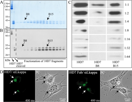
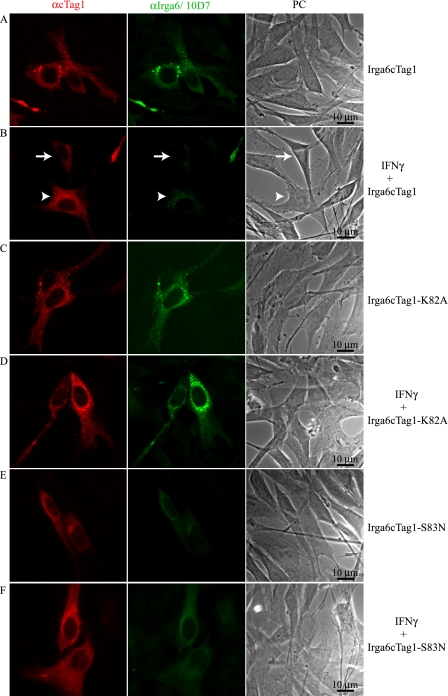
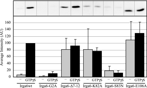
Irga6, a myristoylated, interferon-inducible member of the immunity-related GTPase family, contributes to disease resistance against Toxoplasma gondii in mice. Accumulation of Irga6 on the T. gondii parasitophorous vacuole membrane is associated with vesiculation and ultimately disruption of the vacuolar membrane in a process that requires an intact GTP-binding domain. The role of the GTP-binding domain of Irga6 in pathogen resistance is, however, unclear. We provide evidence that Irga6 in interferon-induced, uninfected cells is predominantly in a GDP-bound state that is maintained by other interferon-induced proteins. However, Irga6 that accumulates on the parasitophorous vacuole membrane after Toxoplasma infection is in the GTP-bound form. We demonstrate that a monoclonal antibody, 10D7, specifically detects GTP-bound Irga6, and we show that the formation of the 10D7 epitope follows from a GTP-dependent conformational transition of the N terminus of Irga6, anticipating an important role of the myristoyl group on Irga6 function in vivo.


Figures

FIGURE 1.
FIGURE 1.
10D7 antibody detects Irga6 at the PVM but not at the ER. gs3T3 fibroblasts were induced with IFNγ for 24 h prior to 2-h infection with T. gondii ME49 strain with a multiplicity of infection of 8. Irga6 protein was labeled with rabbit anti-Irga6 polyclonal serum 165 (red) and with mouse monoclonal anti-Irga6 antibodies 10E7 (A) or 10D7 (B) (green). PC, phase-contrast images. Parasitophorous vacuoles are indicated by the arrowheads. 10D7 detected Irga6 on the PVM efficiently but the cytoplasmic, ER membrane-associated Irga6 at a barely detectable level.
FIGURE 2.
FIGURE 2.
10D7 is a high affinity antibody. Papain-cleaved 10D7 antibody was separated on a Superdex 75 column. Fractions were subjected to SDS-PAGE on 7.5% gels under nonreducing conditions, and protein was detected by colloidal Coomassie staining (A) or Western blot (B) using goat anti-mouse κ light chain and donkey anti-goat horseradish peroxidase antibodies as primary and secondary stage detection reagents, respectively. The apparent molecular weight of native 10D7 IgG on SDS-PAGE was dependent on gel conditions. In relatively short runs in 7.5% gels, 10D7 ran in a complex band pattern below 130 kDa (A and B). However, the same material run longer in a 10% gel behaved rather normally, reaching an average position at or even above 150 kDa (see supplemental Fig. 1). Papain-cleaved fragments were shown to have an apparent molecular mass of ∼40 kDa in the 7.5% gel system (A and B). C, relative affinity of putative Fab fractions B8 and B15 of papain-cleaved 10D7 was estimated by binding to recombinant Irga6 fixed to the nitrocellulose membrane. 10 μg/ml antibodies was considered as 1:1 and further dilutions were made, 1:2, 1:4, 1:8, 1:16, 1:32, and 1:64. A similar dilution series of uncleaved native 10D7 was made as positive controls. Detection of 10D7 and 10D7 fragments was done as in B. Monovalent 10D7 Fab fragment (fraction B15) has an affinity for denatured Irga6 on a Western blot comparable with that of the native 10D7. D, gs3T3 fibroblasts were induced with IFNγ 24 h followed by infection with T. gondii ME49 strain for 2 h. Irga6 was detected with 10 μg/ml of 10D7 antibody (10D7) or 10D7 Fab; as secondary detection reagent, goat anti-mouse κ light chain-fluorescein isothiocyanate (αLkappa) was used. This secondary reagent detects papain-cleaved Fab and intact 10D7 with the same efficiency. The arrows indicate positions of T. gondii vacuoles. PC, phase-contrast images.
FIGURE 3.
FIGURE 3.
In uninfected cells, Irga6 forms GTP-dependent oligomers only in the absence of IFNγ. L929 fibroblasts were simultaneously induced with IFNγ and transfected with Irga6cTag1 24 h before lysis (A), not induced with IFNγ but simultaneously transfected with both Irga6wt and Irga6cTag1 24 h before lysis (B), or either simultaneously ((i + t)) or separately ((i) + (t)) induced with IFNγ and transfected with Irga6cTag1 24 h before lysis (C). Irga6 was immunoprecipitated from lysates with rabbit anti-cTag1 serum. Cells were either preincubated with AlFx and subsequently lysed in the absence of nucleotides (lane 4) or without preincubation lysed in the presence of nucleotides with or without AlFx (lanes 2, 3, 5, and 6). Cells in lane 1 were immunoprecipitated without preincubation and without additives during the immunoprecipitation. Irga6 proteins in immunoprecipitates were detected with 10D7 antibody in a Western blot. A second band above wild-type Irga6cTag1 (marked with a single asterisk) is always found in Western blots of transfected Irga6; the nature of this presumed modification is unknown.
FIGURE 4.
FIGURE 4.
In noninduced cells, 10D7 detects aggregated Irga6 as efficiently as 10E7 antibody. Untreated gs3T3 fibroblasts were transfected with Irga6cTag1 and stained 24 h later with anti-cTag1 polyclonal serum (red) and with either 10E7 (A) or 10D7 (B) monoclonal antibodies (green). PC, phase-contrast images.
FIGURE 5.
FIGURE 5.
Hydrolysis-deficient dominant-negative mutant of Irga6 forms aggregates independently of IFNγ-induced factors and constitutively exposes the 10D7 epitope. Irga6cTag1-wt (A and B), Irga6cTag1-K82A (C and D; functionally dominant negative), and Irga6cTag1-S83N (E and F) constructs were transfected into gs3T3 fibroblasts either in the absence (A, C, and E) or in the presence of IFNγ induction (B, D, and F). Transfected cells were stained 24 h later with 10D7 antibody (green) and anti-cTag1 serum (red). Images were taken with the same exposure time. The arrow and arrowhead in B indicate cells with lower and higher levels of transfected Irga6cTag1 protein, respectively. Treatment with IFNγ markedly attenuates the 10D7 signal on transfected wild-type Irga6cTag1 (B) but has no impact on the 10D7 signal of transfected Irga6cTag1-K82A (C and D). The 10D7 signal is weak on Irga6cTag1-S83N independently of IFNγ treatment (E and F). PC, phase-contrast images.
FIGURE 6.
FIGURE 6.
10D7 epitope is located in the Helix A of Irga6 protein. A, purified, nonmyristoylated recombinant Irga6wt, -Δ7-12, -Δ7-25, -Δ20-25, -F20A, -T21A, -G22A, -Y23A, -F24A, and -K25A proteins were subjected to SDS-PAGE and detected in Western blot with 10E7 and 10D7 antibodies. 10D7 signals were quantified, and values for mutants are given as percentages relative to the Irga6wt signal; B, crystal structure of Irga6 monomer (13), with Helix A, containing the 10D7 epitope, amino acids 20-25, indicated in red. The myristoyl group and the first 12 N-terminal amino acids have not yet been crystallographically resolved. GppNHp and Mg2+ are shown as an atomic stick figure and black sphere, respectively; C, enlarged view of the structure shown in Fig. 6B, looking from below on the 4-helix bundle of which Helix A is a member. The orientations of the side chains of Phe20, Thr21, Tyr23, Phe24, and Lys25 amino acids in the Irga6 structure are shown.
FIGURE 7.
FIGURE 7.
10D7 antibody binds to the GTP-bound form of native cellular Irga6 but not to a myristoylation-deficient mutant. L929 fibroblasts were transfected with Irga6wt, -G2A, -Δ7-12, -K82A, -S83N, or -E106A constructs. Cells were lysed 24 h later in Thesit in the absence or presence of 0.5 mm GTPγS, and Irga6 was immunoprecipitated with 10D7-Protein A-Sepharose beads. Irga6 proteins were detected in Western blot by rabbit anti-Irga6 polyclonal 165 serum. Signals were quantified using ImageQuant TLv2005, and values for immunoprecipitated proteins were normalized to the corresponding lysates. Mean values of at least three independent experiments are shown in the histogram. The 10D7 epitope is dependent on GTPγS in wild type Irga6 but constitutively expressed in functionally dominant negative mutants Irga6-K82A and -E106A. The myristoylation-deficient mutant, Irga6-G2A, cannot express the 10D7 epitope whether GTPγS is present or not. The mutant Irga6-Δ7-12 expresses 10D7 epitope independently of GTPγS.
FIGURE 8.
FIGURE 8.
The myristoyl group, but not the proximal part of Helix A, is important for GTP-dependent Irga6 self-interaction. L929 fibroblasts were transfected with cTag1-tagged (**) and untagged (▸) wild type and mutant Irga6 constructs. The mutants Irga6-G2A, -Δ7-12, -K82A, -S83N, and -E106A were used. For each genotype, cells were transfected simultaneously with tagged and untagged constructs. Cells were lysed in Thesit 24 h later in the absence or presence of GTPγS and immunoprecipitated with anti-cTag1 serum. Irga6 proteins were identified with 10D7 antibody in Western blot. Only the Irga6wt and Irga6-Δ7-12 mutant showed typical GTPγS-dependent co-precipitation (lanes 1 and 3, respectively). The two functionally dominant negative mutants, Irga6-K82A and -E106A, both co-precipitated untagged protein independently of exogenous GTPγS (lane 4 and 6, respectively), whereas Irga6-G2A unexpectedly hardly co-precipitated untagged protein at all (lanes 2). Irga6cTag1-S83N showed no co-immunprecipitation of untagged protein (lanes 5). The upper band in each lane, labeled with a single asterisk, is the unexplained “transfection-specific” band referred to in the legend to Fig. 3.
FIGURE 9.
FIGURE 9.
Model of Irga6 nucleotide-dependent conformational change and membrane interaction. A, the model proposes that regulated GTP binding by wild-type Irga6 initiates a complex conformational change requiring the presence of a “hinge” at residues 7-12 (yellow strand) that releases the myristoyl group (red) from a bound or cryptic configuration and exposes the 10D7 determinant by partial unfolding of Helix A residues 20-25 (green oval). In the absence of the myristoyl group (G2A), the motion is aborted for unknown reasons, and the 10D7 epitope is not exposed. In the Δ7-12 mutant, the critical hinge-like residues are absent, the myristoyl group is constitutively mispositioned, and the 10D7 epitope is constitutively exposed. In the S83N mutant, which cannot bind nucleotides, Irga6 is constitutively in the inactive, closed configuration, and the 10D7 determinant is constitutively not exposed. In the K82A and E106A mutants, Irga6 is constitutively GTP-bound in vivo, and the 10D7 determinant is constitutively exposed. B, in IFNγ-induced cells, the GMS group of 3 IRG proteins favor the GDP-bound inactive state of Irga6, which remains predominantly monomeric in the cytoplasm and at the ER membrane with the 10D7 epitope not exposed. Upon infection, Irga6 is released by an unknown mechanism from GMS control and interacts with the PVM in its GTP-bound form, probably by insertion of the myristoyl group, and exposing the 10D7 epitope. Homooligomerization of GTP-bound Irga6 could increase the avidity of this interaction, stabilizing active Irga6 on the vacuolar membrane.

Similar articles

Cited by

References

    1. Bernstein-Hanley, I., Coers, J., Balsara, Z. R., Taylor, G. A., Starnbach, M. N., and Dietrich, W. F. (2006) Proc. Natl. Acad. Sci. U. S. A. 103 14092-14097 - PMC - PubMed
    1. Butcher, B. A., Greene, R. I., Henry, S. C., Annecharico, K. L., Weinberg, J. B., Denkers, E. Y., Sher, A., and Taylor, G. A. (2005) Infect. Immun. 73 3278-3286 - PMC - PubMed
    1. Collazo, C. M., Yap, G. S., Sempowski, G. D., Lusby, K. C., Tessarollo, L., Woude, G. F., Sher, A., and Taylor, G. A. (2001) J. Exp. Med. 194 181-188 - PMC - PubMed
    1. Feng, C. G., Collazo-Custodio, C. M., Eckhaus, M., Hieny, S., Belkaid, Y., Elkins, K., Jankovic, D., Taylor, G. A., and Sher, A. (2004) J. Immunol. 172 1163-1168 - PubMed
    1. Halonen, S. K., Taylor, G. A., and Weiss, L. M. (2001) Infect. Immun. 69 5573-5576 - PMC - PubMed

Publication types