

Functional connectivity MR imaging reveals cortical functional connectivity in the developing brain
- PMID: 18784212
- PMCID: PMC2583167
- DOI: 10.3174/ajnr.A1256
Functional connectivity MR imaging reveals cortical functional connectivity in the developing brain
Abstract
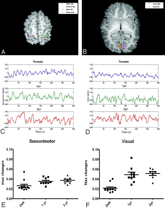
Background and purpose: Unlike conventional functional MR imaging where external sensory/cognitive paradigms are needed to specifically activate different regions of the brain, resting functional connectivity MR imaging acquires images in the absence of cognitive demands (a resting condition) and detects brain regions, which are highly temporally correlated. Therefore, resting functional MR imaging is highly suited for the study of brain functional development in pediatric subjects. This study aimed to determine the temporal and spatial patterns of rfc in healthy pediatric subjects between 2 weeks and 2 years of age.
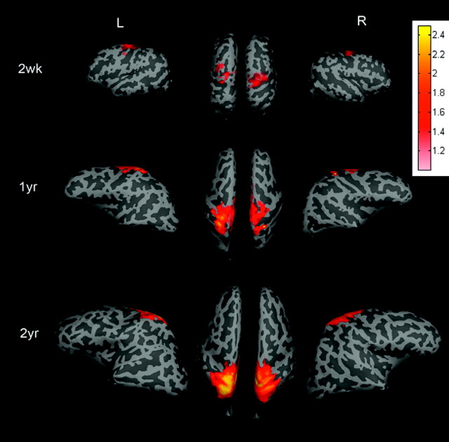
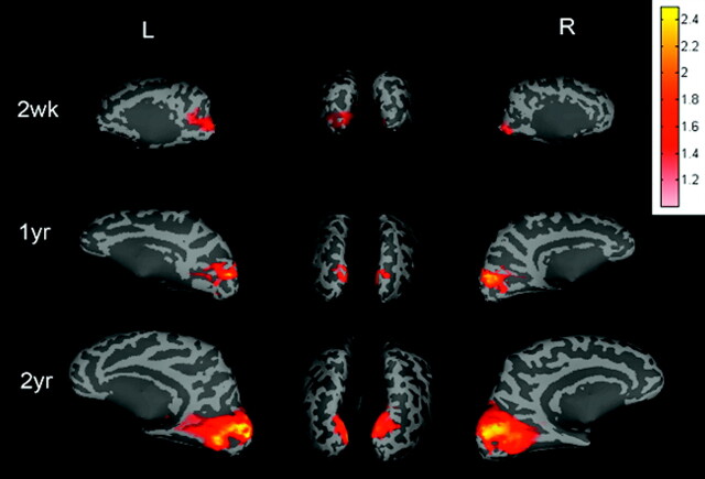
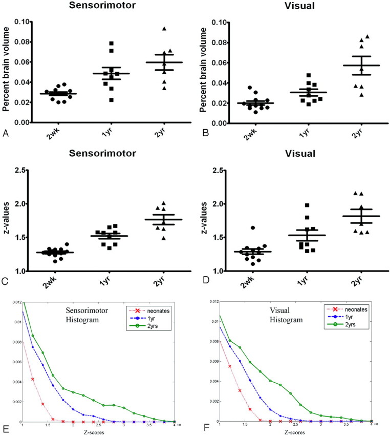
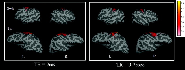
Materials and methods: Rfc studies were performed on 85 children: 38 neonates (2-4 weeks of age), 26 one-year-olds, and 21 two-year-olds. All subjects were imaged while asleep; no sedation was used. Six regions of interest were chosen, including the primary motor, sensory, and visual cortices in each hemisphere. Mean signal intensity of each region of interest was used to perform correlation analysis pixel by pixel throughout the entire brain, identifying regions with high temporal correlation.
Results: Functional connectivity was observed in all subjects in the sensorimotor and visual areas. The percent brain volume exhibiting rfc and the strength of rfc continued to increase from 2 weeks to 2 years. The growth trajectories of the percent brain volume of rfc appeared to differ between the sensorimotor and visual areas, whereas the z-score was similar. The percent brain volume of rfc in the sensorimotor area was significantly larger than that in the visual area for subjects 2 weeks of age (P = .008) and 1-year-olds (P = .017) but not for the 2-year-olds.
Conclusions: These findings suggest that rfc in the sensorimotor precedes that in the visual area from 2 weeks to 1 year but becomes comparable at 2 years. In contrast, the comparable z-score values between the sensorimotor and visual areas for all age groups suggest a disassociation between percent brain volume and the strength of cortical rfc.
Figures
Similar articles
-
Functional connectivity as revealed by spatial independent component analysis of fMRI measurements during rest.Hum Brain Mapp. 2004 Jul;22(3):165-78. doi: 10.1002/hbm.20022. Hum Brain Mapp. 2004. PMID: 15195284 Free PMC article.
-
Disrupted functional connectivity between sub-regions in the sensorimotor areas and cortex in migraine without aura.J Headache Pain. 2020 May 6;21(1):47. doi: 10.1186/s10194-020-01118-1. J Headache Pain. 2020. PMID: 32375638 Free PMC article.
-
Functional connectivity of the sensorimotor area in naturally sleeping infants.Brain Res. 2008 Aug 5;1223:42-9. doi: 10.1016/j.brainres.2008.05.054. Epub 2008 May 28. Brain Res. 2008. PMID: 18599026
-
Longitudinal Study of the Emerging Functional Connectivity Asymmetry of Primary Language Regions during Infancy.J Neurosci. 2016 Oct 19;36(42):10883-10892. doi: 10.1523/JNEUROSCI.3980-15.2016. J Neurosci. 2016. PMID: 27798142 Free PMC article.
-
Neuromagnetic integrated methods tracking human brain mechanisms of sensorimotor areas 'plastic' reorganisation.Brain Res Brain Res Rev. 2000 Sep;33(2-3):131-54. doi: 10.1016/s0169-328x(00)00090-5. Brain Res Brain Res Rev. 2000. PMID: 11011062 Review.
Cited by
-
Longitudinal analysis of neural network development in preterm infants.Cereb Cortex. 2010 Dec;20(12):2852-62. doi: 10.1093/cercor/bhq035. Epub 2010 Mar 17. Cereb Cortex. 2010. PMID: 20237243 Free PMC article.
-
Maturation of large-scale brain systems over the first month of life.Cereb Cortex. 2023 Mar 10;33(6):2788-2803. doi: 10.1093/cercor/bhac242. Cereb Cortex. 2023. PMID: 35750056 Free PMC article.
-
Functional Connectivity Alterations between Networks and Associations with Infant Immune Health within Networks in HIV Infected Children on Early Treatment: A Study at 7 Years.Front Hum Neurosci. 2018 Jan 11;11:635. doi: 10.3389/fnhum.2017.00635. eCollection 2017. Front Hum Neurosci. 2018. PMID: 29375341 Free PMC article.
-
Dynamic causal modelling on infant fNIRS data: A validation study on a simultaneously recorded fNIRS-fMRI dataset.Neuroimage. 2018 Jul 15;175:413-424. doi: 10.1016/j.neuroimage.2018.04.022. Epub 2018 Apr 12. Neuroimage. 2018. PMID: 29655936 Free PMC article.
-
Prenatal cocaine effects on brain structure in early infancy.Neuroimage. 2014 Nov 1;101:114-23. doi: 10.1016/j.neuroimage.2014.06.070. Epub 2014 Jul 3. Neuroimage. 2014. PMID: 24999039 Free PMC article.
References
-
- Biswal BB, Van Kylen J, Hyde JS. Simultaneous assessment of flow and BOLD signals in resting-state functional connectivity maps. NMR Biomed 1997;10:165–70 - PubMed
-
- Biswal B, Hudetz AG, Yetkin FZ, et al. Hypercapnia reversibly suppresses low-frequency fluctuations in the human motor cortex during rest using echo-planar MRI. J Cereb Blood Flow Metab 1997;17:301–08 - PubMed
-
- Lowe MJ, Mock BJ, Sorenson JA. Functional connectivity in single and multislice echoplanar imaging using resting-state fluctuations. Neuroimage 1998;7:119–32 - PubMed
-
- Lowe MJ, Dzemidzic M, Lurito JT, et al. Correlations in low-frequency BOLD fluctuations reflect cortico-cortical connections. Neuroimage 2000;12:582–87 - PubMed
Publication types
MeSH terms
Grants and funding
LinkOut - more resources
Full Text Sources
Medical