. 2008 Nov;19(11):4776-84.
doi: 10.1091/mbc.e08-04-0407. Epub 2008 Sep 10.

Activation of the MAPK module from different spatial locations generates distinct system outputs

Affiliations

Activation of the MAPK module from different spatial locations generates distinct system outputs

Kerry Inder et al. Mol Biol Cell. 2008 Nov.

Abstract

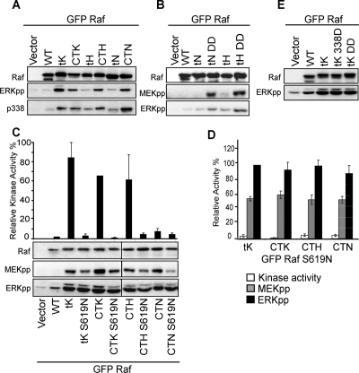
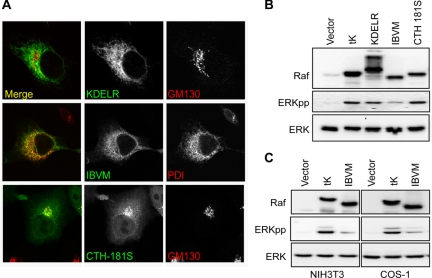
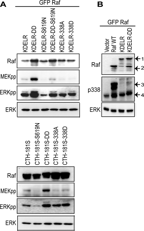
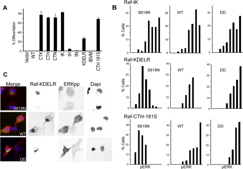
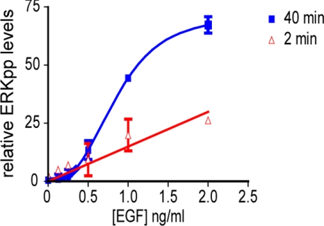
The Ras/Raf/MEK/ERK (MAPK) pathway directs multiple cell fate decisions within a single cell. How different system outputs are generated is unknown. Here we explore whether activating the MAPK module from different membrane environments can rewire system output. We identify two classes of nanoscale environment within the plasma membrane. The first, which corresponds to nanoclusters occupied by GTP-loaded H-, N- or K-Ras, supports Raf activation and amplifies low Raf kinase input to generate a digital ERKpp output. The second class, which corresponds to nanoclusters occupied by GDP-loaded Ras, cannot activate Raf and therefore does not activate the MAPK module, illustrating how lateral segregation on plasma membrane influences signal output. The MAPK module is activated at the Golgi, but in striking contrast to the plasma membrane, ERKpp output is analog. Different modes of Raf activation precisely correlate with these different ERKpp system outputs. Intriguingly, the Golgi contains two distinct membrane environments that generate ERKpp, but only one is competent to drive PC12 cell differentiation. The MAPK module is not activated from the ER. Taken together these data clearly demonstrate that the different nanoscale environments available to Ras generate distinct circuit configurations for the MAPK module, bestowing cells with a simple mechanism to generate multiple system outputs from a single cascade.


Figures

Figure 1.
Figure 1.
Raf-1 targeted to the plasma membrane functions as a digital switch. (A) Raf-CTK, Raf-tK, Raf-CTH, Raf-tN, Raf-CTN, and Raf-tH were transiently expressed in BHK cells and cell lysates prepared after 3 h of serum starvation. Empty vector and wild-type Raf-1 (WT) were used as controls. The Raf-1 Western blot shows equivalent expression of the Raf constructs. Activation of Raf-1 and ERK was assayed with phospho-specific antibodies: pS338-Raf-1 and ERKpp, respectively. (B) BHK cells were transfected with empty vector, wild-type Raf-1 (WT), Raf-tH, Raf-tN, and the cognate constitutively active constructs (DD). Cells were then serum-starved. The Raf-1 Western blot shows equivalent expression of the Raf constructs. Activation of MEK and ERK was assayed with phospho-specific antibodies: MEKpp and ERKpp, respectively. (C) Membrane fractions (P100) prepared from serum-starved BHK cells transiently expressing wild-type Raf-1 (WT), Raf-tK, Raf-CTK, Raf-CTH, or Raf-CTN with and without a kinase mutation (S619N) or from vector transfected control cells were assayed for Raf-1 kinase activity in a coupled MEK/ERK assay. The graph shows mean activity ± SEM (n = 3). Corresponding whole cell lysates were immunoblotted for Raf-1 (to show equivalent Raf expression), MEKpp, and ERKpp. (D) A comparison of Raf kinase activity, MEK activity (MEKpp), and ERK activity (ERKpp) of the kinase reduced (S619N) constructs expressed relative to the cognate wild-type Raf-1 construct (mean ± SEM, n = 3). The results show that each S619N mutant has <5% Raf catalytic activity, ∼50% MEK activity, and 100% ERK activity compared with the wild-type control levels, respectively. Indicative of switch-like signal transmission. (E) BHK cells expressing empty vector, wild-type Raf-1 (WT), Raf-tK, and constitutively active Raf-tK-338D and Raf-tK-DD were serum-starved for 3 h and assayed with Raf-1 and ERKpp antibodies.
Figure 2.
Figure 2.
Raf-1 signaling from the Golgi. (A) Endomembrane-targeted Raf-1 constructs were transiently expressed in BHK cells and colocalized with organelle markers. Raf-KDELR and Raf-IBVM colocalize with GM130 (Golgi marker) and PD1 (ER marker), respectively. Raf-KDELR also shows some degree of ER localization. Raf-CTH-181S localizes exclusively with GM130 to the Golgi. (B) Raf-KDELR, Raf-IBVM, and Raf-CTH-181S were transfected into BHK cells and serum-starved for 3 h. Empty vector and Raf-tK were used as negative and positive controls for ERK activation, respectively. Activation of ERK was assayed in immunoblots of whole cell lysates with phospho-specific antibody ERKpp, and total ERK levels were blotted with anti-ERK 2. (C) ERK activation was assessed from Raf-IBVM expressed in NIH3T3 and COS-1 cells. Empty vector and Raf-tK were used as negative and positive controls. Western blots shows equal loading with ERK antibody and ERK activation with ERKpp phospho-specific antibody.
Figure 3.
Figure 3.
Signaling from the Golgi generates analog signal output. (A) Activation of the MAPK module was assayed in cell lysates prepared from BHK cells transiently expressing Raf-KDELR and Raf-CTH-181S, with different kinase activities constitutively active (DD), kinase-reduced (S619N), both mutations (DD-S619N), or with the point mutations S338A and S338D. The Raf-1 Western blot shows equivalent expression of the Raf constructs and equal total ERK levels. Activation of MEK and ERK was assayed with phospho-specific antibodies: MEKpp and ERKpp, respectively. (B) Raf-KDELR and constitutively active Raf-KDELR-DD transiently expressed in BHK cells show very low levels of Raf S338 phosphorylation detected by immunoblotting with pS338-Raf-1 antisera. Lysates prepared from empty vector and wild-type Raf-1 (WT)-transfected cells are included as negative controls. Equal protein loading is shown by total ERK levels. Arrow 1 points to Raf-KDELR protein, whereas arrow 2 is the degradation product. Arrow 3 corresponds to phosphorylation of the expressed GFP-Raf proteins, and arrow 4 is the phosphorylation of endogenous Raf.
Figure 4.
Figure 4.
Biological outputs from ERKpp are spatially regulated. (A) PC-12 cells transfected with the indicated constructs were assayed for differentiation after 3 d. The graph shows the mean percentage (± SEM, n = 100) of transfected (GFP positive) cells with neurite outgrowth equal to or more than twice the cell body size at 72 h. (B) Serum-starved cells expressing relatively equal Raf-tK, Raf-KDELR, Raf-CTH-181S, and respective S619N and DD mutants were immunostained for pERK. Nuclear pERK was quantitated in single cells (n ≥100) and is displayed in nine bins in the frequency histogram. (C) Representative cells show expression of GFP-Raf-KDELR (WT), GFP-Raf-KDELR-S619N, and GFP-Raf-KDELR-DD, with ERKpp levels detected by Cy3 secondary antibody; DAPI stain of the nucleus; and merged images. Each image displays transfected and nontransfected cells for comparison.
Figure 5.
Figure 5.
Different endogenous signaling dynamics are observed from the plasma membrane versus the Golgi. BHK cells were serum-starved for 3 h and stimulated with EGF at the following concentrations: 0, 0.125, 0.25, 0.5, 1, 2, and 15 ng/ml for 2 min (to assay plasma membrane ERKpp output) and 40 min (to assay Golgi ERKpp output). ERKpp was quantified from Western blots and graphed (mean ± SEM, n = 3). ERKpp values are expressed as a % of the maximal value observed at 2 min with 15 ng/ml EGF. The Golgi ERKpp response rapidly plateaus after 3 ng/ml EGF as shown in the figure, whereas the plasma membrane response continues to increase linearly as shown both here and previously (Tian et al., 2007).

References

    1. Abankwa D., Hanzal-Bayer M., Ariotti N., Plowman S. J., Gorfe A. A., Parton R. G., McCammon J. A., Hancock J. F. A novel switch region regulates H-ras membrane orientation and signal output. EMBO J. 2008;27:727–735. - PMC - PubMed
    1. Apolloni A., Prior I. A., Lindsay M., Parton R. G., Hancock J. F. H-ras but not K-ras traffics to the plasma membrane through the exocytic pathway. Mol. Cell. Biol. 2000;20:2475–2487. - PMC - PubMed
    1. Bivona T. G., Perez De Castro I., Ahearn I. M., Grana T. M., Chiu V. K., Lockyer P. J., Cullen P. J., Pellicer A., Cox A. D., Philips M. R. Phospholipase Cgamma activates Ras on the Golgi apparatus by means of RasGRP1. Nature. 2003;424:694–698. - PubMed
    1. Brunet A., Roux D., Lenormand P., Dowd S., Keyse S., Pouyssegur J. Nuclear translocation of p42/p44 mitogen-activated protein kinase is required for growth factor-induced gene expression and cell cycle entry. EMBO J. 1999;18:664–674. - PMC - PubMed
    1. Chiu V. K., Bivona T., Hach A., Sajous J. B., Silletti J., Wiener H., Johnson R. L., 2nd, Cox A. D., Philips M. R. Ras signalling on the endoplasmic reticulum and the Golgi. Nat. Cell Biol. 2002;4:343–350. - PubMed

Publication types

MeSH terms

Substances