. 2008 Nov;19(11):4826-36.
doi: 10.1091/mbc.e07-05-0442. Epub 2008 Sep 10.

Yeast Golgi-localized, gamma-Ear-containing, ADP-ribosylation factor-binding proteins are but adaptor protein-1 is not required for cell-free transport of membrane proteins from the trans-Golgi network to the prevacuolar compartment

Affiliations

Yeast Golgi-localized, gamma-Ear-containing, ADP-ribosylation factor-binding proteins are but adaptor protein-1 is not required for cell-free transport of membrane proteins from the trans-Golgi network to the prevacuolar compartment

Mohamed E Abazeed et al. Mol Biol Cell. 2008 Nov.

Abstract

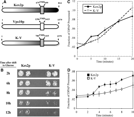
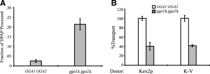
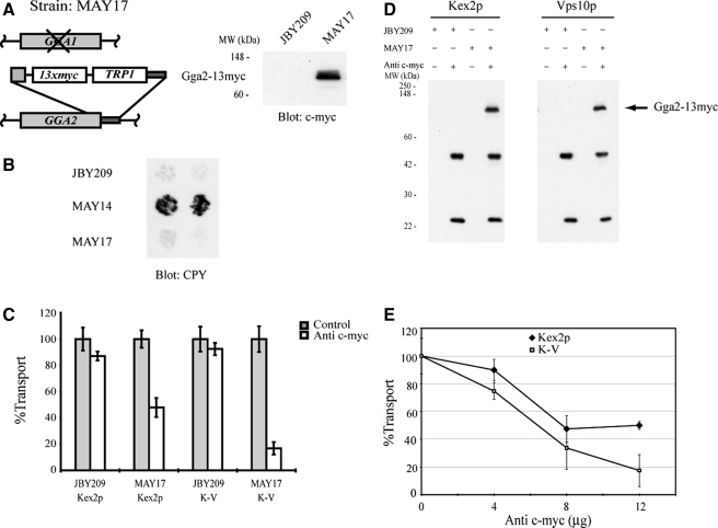
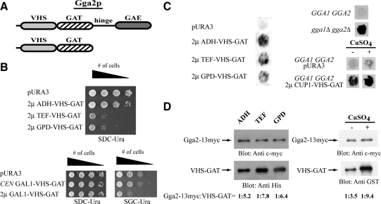
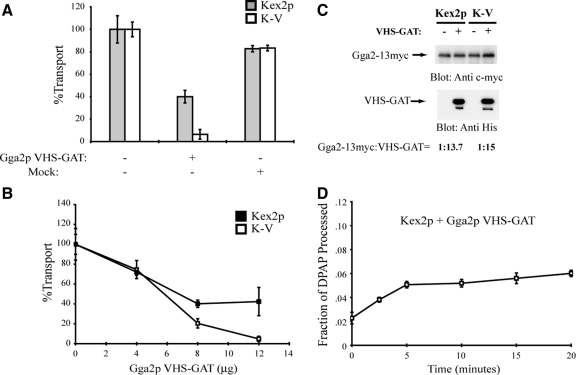
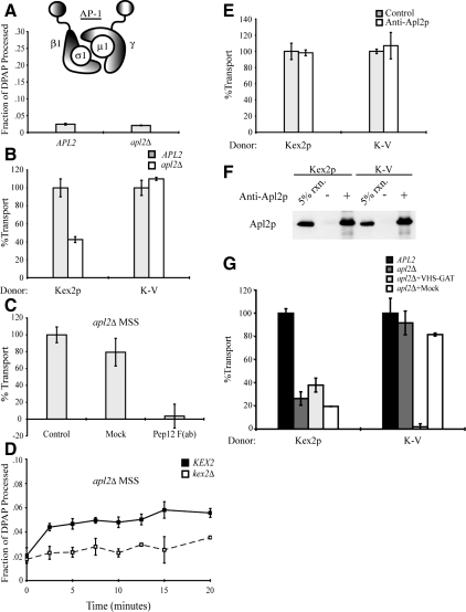
Golgi-localized, gamma-Ear-containing, ADP-ribosylation factor-binding proteins (GGAs) and adaptor protein-1 (AP-1) mediate clathrin-dependent trafficking of transmembrane proteins between the trans-Golgi network (TGN) and endosomes. In yeast, the vacuolar sorting receptor Vps10p follows a direct pathway from the TGN to the late endosome/prevacuolar compartment (PVC), whereas, the processing protease Kex2p partitions between the direct pathway and an indirect pathway through the early endosome. To examine the roles of the Ggas and AP-1 in TGN-PVC transport, we used a cell-free assay that measures delivery to the PVC of either Kex2p or a chimeric protein (K-V), in which the Vps10p cytosolic tail replaces the Kex2p tail. Either antibody inhibition or dominant-negative Gga2p completely blocked K-V transport but only partially blocked Kex2p transport. Deletion of APL2, encoding the beta subunit of AP-1, did not affect K-V transport but partially blocked Kex2p transport. Residual Kex2p transport seen with apl2Delta membranes was insensitive to dominant-negative Gga2p, suggesting that the apl2Delta mutation causes Kex2p to localize to a compartment that precludes Gga-dependent trafficking. These results suggest that yeast Ggas facilitate the specific and direct delivery of Vps10p and Kex2p from the TGN to the PVC and that AP-1 modulates Kex2p trafficking through a distinct pathway, presumably involving the early endosome.


Figures

Figure 1.
Figure 1.
The C-tails of Kex2p and Vps10p regulate the rate of transport between the TGN and the PVC. (A) Schematic depiction of the domain composition of Kex2p, Vps10p, and the chimeric protein K-V. The numbers above each construct designate the N- and C-terminal amino acids of lumenal, transmembrane, and cytosolic domains. TMD, transmembrane domain. (B) KRY24-2D (MATα kex2) cells expressing Kex2p or K-V under the control of the GAL1 promoter were shifted from galactose to glucose for the indicated times before testing mating competence as described in Materials and Methods. (C and D) Cell-free transport reactions were conducted as described in Materials and Methods by using MSS isolated from JBY209 transformed with pKEX2 (Kex2p; C and D) or transformed with pKEX2-VPS10 (K-V; C and D). Reactions were incubated at 30°C for the indicated times.
Figure 2.
Figure 2.
Deletion of the GGAs results in increased PSHA processing and reduced cell-free transport of Kex2p and K-V. (A) PSHA is cleaved in vivo as a result of gga1 gga2 disruption. MSS samples prepared from JBY209 (kex2Δ GGA1+ GGA2+) and MAY8 (kex2Δ gga1Δ gga2Δ), both transformed with pPSHA, were incubated under transport reaction conditions and then subjected to IP with anti-HA antibody and mock IP in the presence of 1% Triton X-100. Supernatants were assayed for DPAP activity by using Ala-ProAMC (Brickner et al., 2001). The fraction of PSHA cleaved in vivo (i.e., lacking the HA epitopes) is the ratio of DPAP activity in the anti-HA IP to that in the mock IP. (B) Donor MSS were prepared from JBY209 (GGA1+ GGA2+) and MAY4 (gga1Δ gga2Δ) expressing Kex2p or K-V. Acceptor MSS were prepared from JBY209 (GGA1+ GGA2+) expressing PSHA. Cell-free transport reactions were then carried out (20 min at 30°C). Extents of transport observed for controls (GGA1+ GGA2+) were 9.2% JBY209 (Kex2p) and 10.3% JBY209 (K-V), adjusted to 100% in the figure. Experimental values (gga1Δ gga2Δ) are shown as percentage of the control values.
Figure 3.
Figure 3.
Gga2p is required for cell-free TGN-to-PVC transport of Kex2p and K-V. (A) Schematic depiction of MAY17 construction (see Materials and Methods) and expression of Gga2p-myc. Whole-cell lysates were prepared from JBY209 (GGA1 GGA2) and MAY17 (gga1 GGA2::13xmyc). Proteins were resolved by SDS-PAGE (10% acrylamide body gel) and analyzed by immunoblotting using anti-c-myc. The position of Gga2p-myc and molecular mass size markers are indicated. (B) GGA2::13myc expresses Gga2-13myc and functionally replaces Gga2p. JBY209 (GGA1 GGA2), MAY14 (gga1 gga2), and MAY17 (gga1 GGA2::13myc) were analyzed for the Vps phenotype (CPY secretion; see Materials and Methods). (C) MSS was prepared from strains JBY209 (GGA1 GGA2) and MAY17 (gga1 GGA2::13myc) expressing Kex2p, K-V or PSHA. Donor and acceptor MSS samples were combined and preincubated with 12 μg of anti-c-myc for 1 h on ice. Cell-free transport reactions were then carried out (20 min at 30°C). Extents of transport observed for controls (no anti-c-myc) were 11.3%, JBY209 (Kex2p); 13.8%, MAY17 (Kex2p); 10.4%, JBY209 (K-V); and 11.9%, MAY17 (K-V), adjusted to 100% in the figure. Experimental values (+ anti-c-myc) are shown as percentage of the control values. (D) Anti-c-myc recognizes Gga2p-myc during the course of the reaction. An aliquot of each reaction shown in C was incubated with protein A-Sepharose for 1 h at 4°C. Protein A-Sepharose pellets were subsequently washed and solubilized in SDS-PAGE sample buffer. Eluted samples were resolved by SDS-PAGE and analyzed by immunoblotting using anti-c-myc. (E) Titration of anti-c-myc in transport reactions. Donor and acceptor MSS samples were combined and preincubated with the indicated amounts of anti-c-myc for 1 h on ice. Transport reactions were then carried out (20 min at 30°C). Data are expressed as percentage of control reactions as described in (C). Extents of transport in control (no anti-c-myc) reactions were 11.2% MAY17 (Kex2p) and 11.9% MAY17 (K-V).
Figure 4.
Figure 4.
Gga2p VHS-GAT functions as a dominant negative in vivo. (A) Schematic representation of Gga2p and Gga2p VHS-GAT. (B) High level expression of Gga2p VHS-GAT causes slow growth. Strain CRY2 was transformed with single-copy CEN vectors (p416) or high-copy 2μ vectors (p426) vectors expressing Gga2p VHS-GAT (aa 1-336) under the indicated promoters. Transformed strains were pronged at decreasing cell densities on SDC-Ura or SGC-Ura plates and tested for growth at 30°C for ∼2 d. (C) High level expression of Gga2p VHS-GAT causes secretion of CPY. Strain CRY2 was transformed with the indicated vectors and transformants were tested for the Vps phenotype (CPY secretion; see Materials and Methods). Also, strains CRY2 (GGA1 GGA2), MAY14 (gga1Δ gga2Δ), CRY2 (pURA3), and CRY2 (2 μ CUP1-VHS-GAT) were tested for the Vps phenotype (CPY secretion; see Materials and Methods). Filters were shifted to SDC-Ura or SDC-Ura supplemented containing 100 μM CuSO4 for 6 h before immunoblot analysis with anti-CPY antibody. (D) Whole-cell lysates were prepared from MAY17 (gga1 GGA2::13xmyc) expressing Gga2p VHS-GAT under ADH, TEF, GPD, or CUP1 promoters. Proteins were resolved by SDS-PAGE (10% acrylamide body gel) and analyzed by immunoblotting using anti-c-myc and anti-His or anti-GST. Quantification of ratios of Gga2-13myc:VHS-GAT was performed as described in Materials and Methods.
Figure 5.
Figure 5.
Purified Gga2p VHS-GAT inhibits cell-free TGN-to-PVC transport reactions containing Kex2p and K-V donor membranes. (A) MSS was prepared from strain JBY209 expressing Kex2p, K-V, or PSHA. Donor and acceptor MSS samples were combined and preincubated with 12 μg of purified Gga2p VHS-GAT, with a mock-purified sample (equivalent volume) or with no addition (control), for 1 h on ice. Cell-free transport reactions were then carried out (20 min at 30°C). Extents of transport observed for the controls (no addition) were 10.8% JBY209 (Kex2p), 11.7% JBY209 (K-V), adjusted to 100% in the figure. Reactions containing Gga2p VHS-GAT or mock addition are shown as percentage of the control values. (B) Titration of Gga2p VHS-GAT in transport reactions. Donor and acceptor MSS samples were combined and preincubated with the indicated amounts of purified Gga2p VHS-GAT or with no addition (control) for 1 h on ice. Transport reactions were then carried out (20 min at 30°C). Data are expressed as percentage of control reactions as described in A. Extents of transport in control (no addition) reactions were 12.3% JBY209 (Kex2p), 9.9% JBY209 (K-V). (C) MSS was prepared from strain MAY17 (gga1 GGA2::13xmyc) expressing Kex2p, K-V, or PSHA. Donor and acceptor MSS samples were combined and incubated with 12 μg of purified Gga2p VHS-GAT with a C-terminal 6X His tag or with no addition. Proteins were resolved by SDS-PAGE (10% acrylamide body gel) and analyzed by immunoblotting using anti-c-myc and anti-His. Quantification of ratios of Gga2p:VHS-GAT was performed as described in Materials and Methods. (D) Donor and acceptor MSS from strain JBY209 expressing Kex2p or PSHA were combined and preincubated with Gga2p VHS-GAT as described in A. A time course of cell-free transport was then measured as Figure 1C.
Figure 6.
Figure 6.
AP-1 is not required for cell-free TGN-to-PVC transport of Kex2p and K-V, but it is required for localization of Kex2p to the Gga-dependent donor compartment. (A) PSHA is not cleaved in vivo as a result of AP-1 disruption. MSS samples prepared from JBY209 (kex2Δ APL2+) and MAY8 (kex2Δ apl2Δ), both transformed with pPSHA, were incubated under transport reaction conditions then subjected to IP with anti-HA antibody and mock IP in the presence of 1% Triton X-100, and supernatants were assayed for DPAP activity by using Ala-ProAMC (Brickner et al., 2001). The fraction of PSHA cleaved in vivo (i.e., lacking the HA epitopes) is the ratio of DPAP activity in the anti-HA IP to that in the mock IP. (B) MSS were prepared from JBY209 (APL2+) and MAY8 (apl2Δ) expressing Kex2p, K-V, or PSHA. Cell-free transport reactions were then carried out (20 min at 30°C). Extents of transport observed for controls (APL2+) were 10.5% JBY209 (Kex2p), 9.7% JBY209 (K-V), adjusted to 100% in the figure. Experimental values (apl2Δ) are shown as percentage of the control values. (C) Residual Kex2p transport in apl2Δ MSS is dependent on Pep12p. Anti-Pep12p was subjected to papain cleavage to create F(ab) fragments against the cytosolic domain of Pep12p. A mock papain cleavage reaction (Mock) was performed to control for potential papain-dependent inhibition. Kex2p and PSHA MSS from strain MAY8 (apl2Δ) were combined and preincubated with 3.5 μg of anti-Pep12p F(ab), with a mock-purified sample (equivalent volume) or with no addition (control), for 1 h on ice. Cell-free transport reactions were then carried out (20 min at 30°C). Extent of transport observed for control [no Mock or anti-Pep12p F(ab)] was 6.2%, adjusted to 100% in the figure. Experimental values [Mock and anti-Pep12p F(ab)] are shown as percentage of the control values. (D) kex2Δ, KEX2+, or PSHA MSS from strain MAY8 (apl2Δ) were combined and a time course of cell-free transport was then measured as Figure 1C. (E) MSS was prepared from strain JBY209 (APL2) expressing Kex2p, K-V or PSHA. Donor and acceptor MSS samples were combined and preincubated with 14 μg of anti-Apl2p antibody or no antibody (control) for 1 h on ice. Cell-free transport reactions were then carried out (20 min at 30°C). Extents of transport observed for controls (no anti-Apl2p) were 12.4% JBY209 (Kex2p), 11.8% JBY209 (K-V), adjusted to 100% in the figure. Experimental values (+ anti-Apl2p) are shown as percentage of the control values. (F) Affinity-purified anti-Apl2p recognizes Apl2p during the course of the reaction. An aliquot of each reaction shown in E was incubated with protein A-Sepharose for 1 h at 4°C. Protein A-Sepharose pellets were subsequently washed and solubilized in SDS-PAGE sample buffer. Eluted samples were resolved by SDS-PAGE and analyzed by immunoblotting using anti-Apl2p. (G) Residual Kex2p transport in reactions containing donor and acceptor MSS from MAY8 (apl2Δ) is resistant to inhibition by purified Gga2 VHS-GAT. MSS were prepared from JBY209 (APL2+) and MAY8 (apl2Δ) expressing Kex2p, K-V, or PSHA. Donor and acceptor MSS samples from MAY8 (apl2Δ) were combined and preincubated with 12 μg of purified Gga2p VHS-GAT, with a mock-purified sample (equivalent volume) or with no addition, for 1 h on ice. Donor and acceptor MSS samples from JBY209 (APL2+) were combined and preincubated with no addition (control). Cell-free transport reactions were then carried out (20 min at 30°C). Extents of transport observed for the controls (APL2+, no addition) were 9.9% JBY209 (Kex2p), 10.1% JBY209 (K-V), adjusted to 100% in the figure. Reactions containing MSS from apl2Δ are shown as percentage of the control values.

References

    1. Abazeed M. E., Blanchette J. M., Fuller R. S. Cell-free transport from the trans-Golgi network to late endosome requires factors involved in formation and consumption of clathrin-coated vesicles. J. Biol. Chem. 2005;280:4442–4450. - PubMed
    1. Becherer K. A., Rieder S. E., Emr S. D., Jones E. W. Novel syntaxin homologue, Pep12p, required for the sorting of lumenal hydrolases to the lysosome-like vacuole in yeast. Mol. Biol. Cell. 1996;7:579–594. - PMC - PubMed
    1. Black M. W., Pelham H. R. A selective transport route from Golgi to late endosomes that requires the yeast GGA proteins. J. Cell Biol. 2000;151:587–600. - PMC - PubMed
    1. Blanchette J. M., Abazeed M. E., Fuller R. S. Cell-free reconstitution of transport from the trans-Golgi network to the late endosome/prevacuolar compartment. J. Biol. Chem. 2004;279:48767–48773. - PubMed
    1. Bonifacino J. S. The GGA proteins: adaptors on the move. Nat. Rev. Mol. Cell Biol. 2004;5:23–32. - PubMed

Publication types

MeSH terms

Substances