

doi: 10.1523/JNEUROSCI.2494-08.2008.
Specificity of immunoreactions: the importance of testing specificity in each method
Affiliations
- PMID: 18784286
- PMCID: PMC2629537
- DOI: 10.1523/JNEUROSCI.2494-08.2008
Item in Clipboard
Specificity of immunoreactions: the importance of testing specificity in each method
J Neurosci.
.
No abstract available
Figures
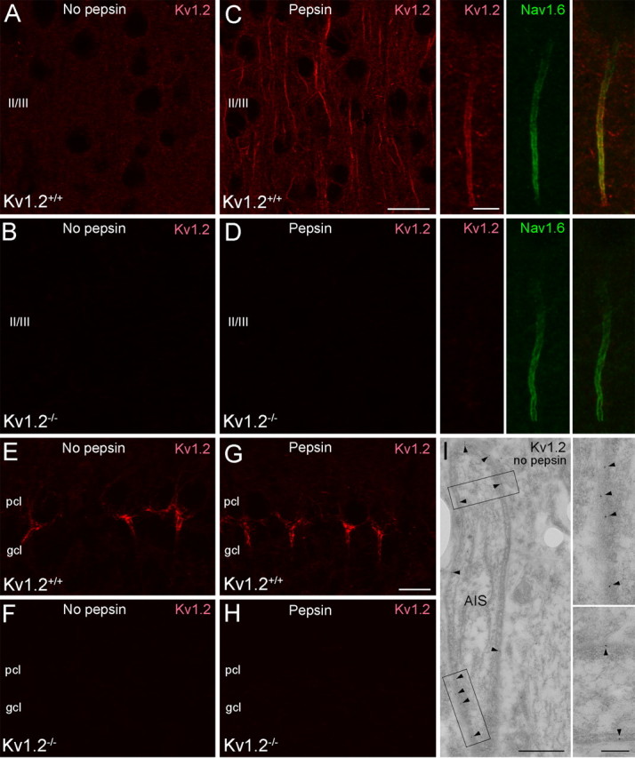
The effect of antigen presentation and subcellular environment on the detectability of the Kv1.2 subunit. A, B, Conventional immunofluorescent reaction reveals a weak neuropil labeling for the Kv1.2 subunit in the neocortex of Kv1.2+/+ mice (A), which completely disappears in Kv1.2−/− mice (B). C, In pepsin-treated control neocortical sections, in addition to the neuropil labeling, an intense Kv1.2 subunit immunolabeling appears in AISs. C, Inset, Intense Kv1.2 subunit immunolabeling outlines the plasma membrane of an Nav1.6-subunit-immunopositive AIS. D, In pepsin-treated Kv1.2−/− neocortical sections, all immunoreactivity disappeared. E, G, The Kv1.2 subunit immunoreactivity of axonal plexus of cerebellar basket cells (pinceau) is revealed with (G) or without (E) pepsin treatment. F, H, The labeling disappears in Kv1.2−/− mice. I, An electron micrograph shows postembedding Kv1.2 subunit immunogold reaction in the neocortex. Immunogold particles (arrowhead) associated with the plasma membrane of an AIS could be detected without pepsin treatment. Insets show the boxed areas of the main panel at a higher magnification. Scale bars: (in C) A–D, 20 μm; C, D, inset, 5 μm; (in G) E–H, 20 μm; I, 500 nm; I, inset, 100 nm.
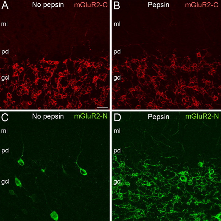
The effect of different antigen presentation depends on the location of the epitope. A, B, A similar axosomatodendritic labeling pattern was detected in cerebellar Golgi cells with (B) or without (A) pepsin treatment when a C-terminal anti-mGluR2 antibody (mGluR2-C) was used. C, Immunofluorescent reaction with an anti-mGluR2 antibody raised against an N-terminal epitope (mGluR2-N) reveals a somatic and proximal dendritic cytoplasmic labeling of cerebellar Golgi cells. D, After pepsin treatment, the mGluR2-N antibody could access the epitopes in all subcellular locations, resulting in the well known axosomatodendritic immunolabeling pattern. All panels are at the same magnification. Scale bar, 20 μm
References
-
- Farrant M, Nusser Z. Variations on an inhibitory theme: phasic and tonic activation of GABA(A) receptors. Nat Rev Neurosci. 2005;6:215–229. - PubMed
-
- Jones A, Korpi ER, McKernan RM, Pelz R, Nusser Z, Mäkelä R, Mellor JR, Pollard S, Bahn S, Stephenson FA, Randall AD, Sieghart W, Somogyi P, Smith AJ, Wisden W. Ligand-gated ion channel subunit partnerships: GABAA receptor α6 subunit gene inactivation inhibits δ subunit expression. J Neurosci. 1997;17:1350–1362. - PMC - PubMed
-
- Ohishi H, Ogawa-Meguro R, Shigemoto R, Kaneko T, Nakanishi S, Mizuno N. Immunohistochemical localization of metabotropic glutamate receptors, mGluR2 and mGluR3, in rat cerebellar cortex. Neuron. 1994;13:55–66. - PubMed
-
- Pool CW, Buijs RM. Antigen identity in immunocytochemistry. In: Van Leeuwen FW, Buijs RM, Pool CW, Pach O, editors. Molecular neuroanatomy. Vol 3. Amsterdam: Elsevier; 1988.
Publication types
MeSH terms
Grants and funding
LinkOut - more resources
Full Text Sources
Other Literature Sources
Molecular Biology Databases