. 2008 Sep 10;28(37):9173-82.
doi: 10.1523/JNEUROSCI.2783-08.2008.

A novel Na+ channel splice form contributes to the regulation of an androgen-dependent social signal

Affiliations

A novel Na+ channel splice form contributes to the regulation of an androgen-dependent social signal

He Liu et al. J Neurosci. .

Abstract

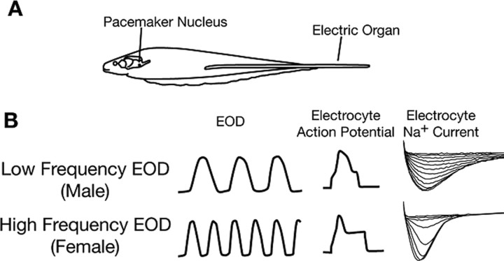
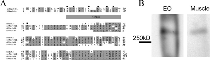
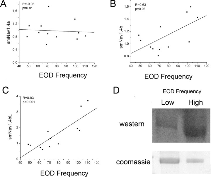
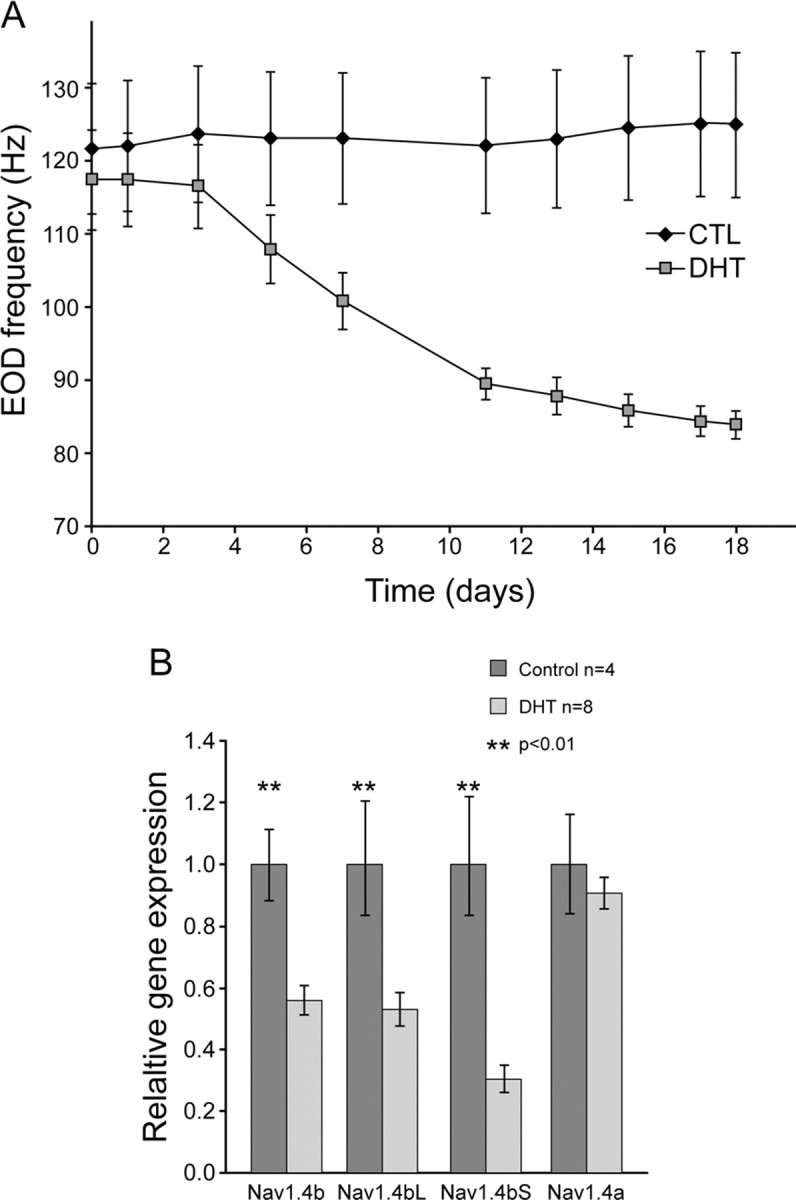
Na(+) channels are often spliced but little is known about the functional consequences of splicing. We have been studying the regulation of Na(+) current inactivation in an electric fish model in which systematic variation in the rate of inactivation of the electric organ Na(+) current shapes the electric organ discharge (EOD), a sexually dimorphic, androgen-sensitive communication signal. Here, we examine the relationship between an Na(+) channel (Na(v)1.4b), which has two splice forms, and the waveform of the EOD. One splice form (Na(v)1.4bL) possesses a novel first exon that encodes a 51 aa N-terminal extension. This is the first report of an Na(+) channel with alternative splicing in the N terminal. This N terminal is present in zebrafish suggesting its general importance in regulating Na(+) currents in teleosts. The extended N terminal significantly speeds fast inactivation, shifts steady-state inactivation, and dramatically enhances recovery from inactivation, essentially fulfilling the functions of a beta subunit. Both splice forms are equally expressed in muscle in electric fish and zebrafish but Na(v)1.4bL is the dominant form in the electric organ implying electric organ-specific transcriptional regulation. Transcript abundance of Na(v)1.4bL in the electric organ is positively correlated with EOD frequency and lowered by androgens. Thus, shaping of the EOD waveform involves the androgenic regulation of a rapidly inactivating splice form of an Na(+) channel. Our results emphasize the role of splicing in the regulation of a vertebrate Na(+) channel and its contribution to a known behavior.


Figures

Figure 1.
Figure 1.
Relationship between the EOD and Na+ current inactivation kinetics. A, EOD frequency is determined by the firing rate of pacemaker neurons in the pacemaker nucleus. The ∼50 pacemaker neurons are electrotonically coupled and fire simultaneously. Each action potential travels down to spinal motor neurons (not depicted) and these innervate the electrocytes, the cells of the electric organ. There is a 1:1 relationship between the firing rate of the pacemaker neurons and the EOD frequency as each descending action potential initiates a pulse from the electric organ. B, The duration of each pulse of the EOD is determined by the membrane properties of the electrocytes. Males generate long-duration pulses at low frequencies (top left), females generate short-duration pulses at high frequencies (bottom left). EOD pulse duration depends on the duration of the electrocyte action potentials (note stylized action potential riding on current injection, center), whose duration is mostly determined by the rate of inactivation of the electrocyte Na+ current (right). [This figure is reprinted from Liu et al. (2007), their Fig. 1].
Figure 2.
Figure 2.
Identification of a novel Nav1.4 with an extended N terminus in Sternopygus. A, RT-PCR showed alternative splicing patterns in the electric organ (E) and skeletal muscle (M) of Sternopygus macrurus and in the skeletal muscle of the zebrafish (Danio rerio) and the rat (Rattus norvegicus). The DNA ladder (L) is in base pairs. B, Schematic illustration of the alternative splice pattern of Nav1.4 in the three species. Each block represents an exon with the assigned name marked above. The size of each intron and exon is marked below if known.
Figure 3.
Figure 3.
Extended N terminus of smNav1.4bL. A, Alignment of translated N-terminal amino acid sequences of Nav1.4b in Sternopygus macrurus (smNav1.4bL) and the zebrafish Danio rerio (drNav1.4bL), the other EO-expressing Na+ channel gene from S. macrurus (smNav1.4a), and their human ortholog (hNav1.4). Residues with similar functionality are shaded. Round dots mark the positively charged residues in the predicted helical region of the extended N terminus of smNav1.4bL. A square marks the proline residue immediately after the helix and mutated in this study. B, Immunoprecipitation of Na+ channel with N-terminal specific antibody followed by labeling with a pan-Na+ channel antibody on Western blots confirms the translation of the extended N terminus and the expression of the long splice form of smNav1.4b in Sternopygus muscle and EO.
Figure 4.
Figure 4.
Properties of hNav1.4, smNav1.4bS, and smNav1.4bL expressed in Xenopus oocytes. A, Averaged peak current traces show that smNav1.4bL inactivates faster than smNav1.4bS and both are faster than their human ortholog hNav1.4. B, The three channels show no significant difference in voltage-dependent activation. C, smNav1.4bL shows a leftward shift of steady-state inactivation compared with the other two channels. Results of hNav1.4 and smNav1.4bS in this figure and Figure 5A–D were published previously by Liu et al. (2007) and replotted with smNav1.4bL for comparison.
Figure 5.
Figure 5.
β1 subunits differentially modulate smNav1.4bL and smNav1.4bS. A, B, Both alternatively spliced β1 subunits speed up the inactivation (A) and negatively shift the steady-state voltage dependence of inactivation (B) of smNav1.4bS. C, The β1L subunit slightly but significantly speeds up the inactivation of smNav1.4bL, whereas β1S has no effect. D, β1 subunits do not shift the voltage dependence of steady-state inactivation of smNav1.4bL. E, smNav1.4bS shows extraordinarily slow recovery from inactivation, but the recovery is much faster when coexpressed with a β1 subunit (for clarity, only β1L is illustrated). F, In contrast, smNav1.4bL shows fast recovery from inactivation with or without a β1 subunit.
Figure 6.
Figure 6.
Deletion of a proline in the extended N terminal partially abolishes the difference between smNav1.4bL and smNav1.4bS. A, Representative current traces show that the proline deletion slows the inactivation of smNav1.4bL. B, The three channels show no significant difference in voltage-dependent activation. C, The deletion of the proline shifts the voltage-dependent inactivation toward the direction of smNav1.4bS. D, The deletion of proline does not significantly change the recovery from inactivation.
Figure 7.
Figure 7.
Semiquantitative RT-PCR reveals the expression profiles of Na+ channels in muscle and electric organ of Sternopygus. A, RT-PCR with primers specific to smNav1.4bL or smNav1.4bS. The same amount of total RNA from the electric organ and skeletal muscle was loaded as template. One-step RT-PCRs were set up identically but stopped after different numbers of cycles. Comparison is made between tissue types for each splice form, showing that smNav1.4bL (L) has a comparable abundance in muscle and electric organ whereas smNav1.4bS (S) is very low in the electric organ. B, smNav1.4a and smNav1.4b have a comparable abundance in the electric organ of midfrequency EOD (∼90 Hz) fish. One-step RT-PCRs with primers in conserved regions were set up identically but stopped after different numbers of cycles. Products with expected sizes (519 bp, top band, from smNav1.4a and 450bp, bottom band, from smNav1.4b) were separated and shown in a 2% agarose gel. Comparisons made between the two genes show that they have comparable abundance in a sample of fish with a midrange EOD frequency.
Figure 8.
Figure 8.
The expression of smNav1.4a, smNav1.4b, and the smNav1.4bL splice variant versus EOD frequency. Measurements are by RT-PCR and normalized to 18S RNA. A, mRNA level of smNav1.4a shows no significant correlation with each individual's EOD frequency. B, C, mRNA levels of smNav1.4b and its major splice variant smNav1.4bL are significantly higher in fish with high than with low EOD frequencies and positively correlated with EOD frequency. D, Protein samples from the EO of fish with high or low EOD frequencies were immunoprecipitated by a pan Na+ channel antibody, loaded on SDS-PAGE gels, and stained with either Coomassie blue (bottom) or blotted with the antibody against smNav1.4bL extended N terminus (top, slight difference in apparent molecular weight because of “smiling” on the gel). The comparison shows that a greater proportion of the total Na+ channel protein is Nav1.4bL in fish with higher EOD frequencies. CTL, Control.
Figure 9.
Figure 9.
DHT lowered the EOD frequency and the expression of smNav1.4b. A, DHT implants lowered the EOD frequency over a period of 18 d. The EOD frequency was significantly different by 7 d after the DHT implant. B, Real-time quantitative RT-PCR shows that the expression of smNav1.4b and its splice variant Nav1.4bL are lowered by DHT implants. There is no significant change in the expression of smNav1.4a. [The data in A have been published previously in and are adapted from Liu et al. (2007), their Fig. 7A]. CTL, Control.
Figure 10.
Figure 10.
A hypothesis for molecular regulation of the rate of inactivation of the Na+ current in the Sternopygus electrocyte. A, Schematic illustration of how mRNA levels of smNav1.4a, smNav1.4bL, and the β1 subunit vary with systematic variation in EOD waveform and electrocyte Na+ current. B, We hypothesize that smNav1.4a inactivates slowly. In this study, we show that smNav1.4bL activates rapidly. We hypothesize that two factors regulate the rate of inactivation: (1) as the ratio of smNav1.4bL to smNav1.4a increases, the average rate of inactivation of the population of Na+ channels will be faster; (2) because β1 subunits speed the rate of inactivation of Na+ currents, the increase in levels of β1 will speed both currents, especially Nav1.4a.

Similar articles

Cited by

References

    1. Akopian AN, Okuse K, Souslova V, England S, Ogata N, Wood JN. Trans-splicing of a voltage-gated sodium channel is regulated by nerve growth factor. FEBS Lett. 1999;445:177–182. - PubMed
    1. Becker MN, Brenner R, Atkinson NS. Tissue-specific expression of a Drosophila calcium-activated potassium channel. J Neurosci. 1995;15:6250–6259. - PMC - PubMed
    1. Blumenstein Y, Kanevsky N, Sahar G, Barzilai R, Ivanina T, Dascal N. A novel long N-terminal isoform of human L-type Ca2+ channel is up-regulated by protein kinase C. J Biol Chem. 2002;277:3419–3423. - PubMed
    1. Bohm RA, Wang B, Brenner R, Atkinson NS. Transcriptional control of Ca(2+)-activated K(+) channel expression: identification of a second, evolutionarily conserved, neuronal promoter. J Exp Biol. 2000;203:693–704. - PubMed
    1. Chew LJ, Gallo V. Regulation of ion channel expression in neural cells by hormones and growth factors. Mol Neurobiol. 1998;18:175–225. - PubMed

Publication types

MeSH terms

LinkOut - more resources