. 2008 Sep 10;28(37):9194-204.
doi: 10.1523/JNEUROSCI.3314-07.2008.

Noggin expands neural stem cells in the adult hippocampus

Affiliations

Noggin expands neural stem cells in the adult hippocampus

Michael A Bonaguidi et al. J Neurosci. .

Abstract

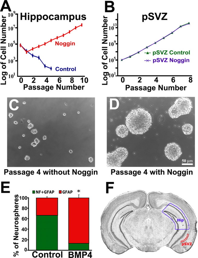
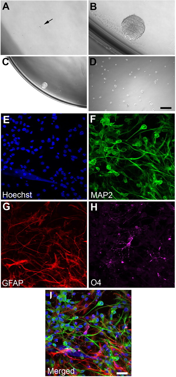
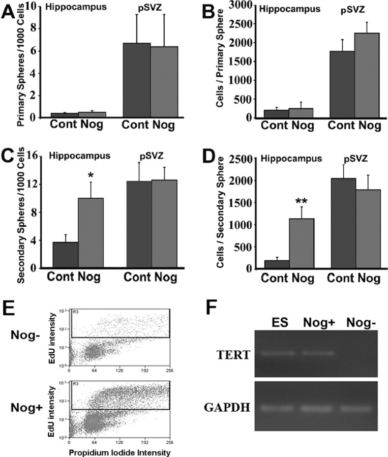
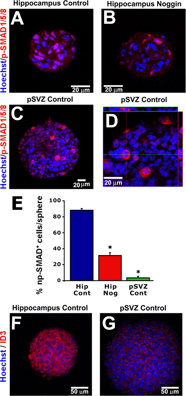
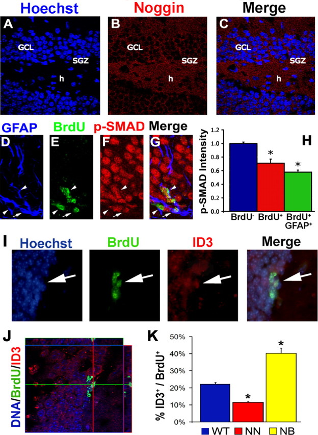
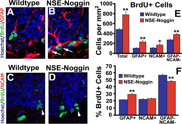
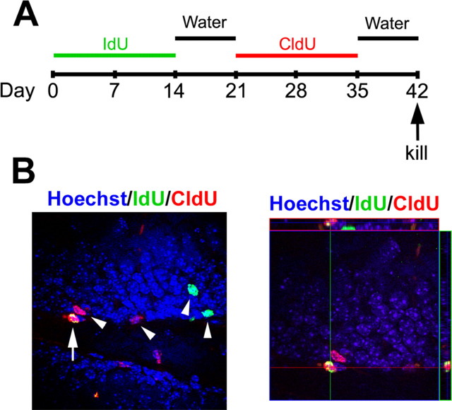
New neurons are added to the adult hippocampus throughout life and contribute to cognitive functions, including learning and memory. It remains unclear whether ongoing neurogenesis arises from self-renewing neural stem cells (NSCs) or from multipotential progenitor cells that cannot self-renew in the hippocampus. This is primarily based on observations that neural precursors derived from the subventricular zone (SVZ) can be passaged long term, whereas hippocampal subgranular zone (SGZ) precursors are rapidly depleted by passaging. We demonstrate here that high levels of bone morphogenetic protein (BMP) signaling occur in hippocampal but not SVZ precursors in vitro, and blocking BMP signaling with Noggin is sufficient to foster hippocampal cell self-renewal, proliferation, and multipotentiality using single-cell clonal analysis. Moreover, NSC maintenance requires continual Noggin exposure, which implicates BMPs as crucial regulators of NSC aging. In vivo, Noggin is expressed in the adult dentate gyrus and limits BMP signaling in proliferative cells of the SGZ. Transgenic Noggin overexpression in the SGZ increases multiple precursor cell populations but proportionally increases the glial fibrillary acidic protein-positive cell population at the expense of other precursors, suggesting that Noggin acts on NSCs in vivo. To confirm this, we used a dual thymidine analog paradigm to repeatedly label slowly dividing cells over a long duration. We find that small populations of label-retaining cells exist in the SGZ and that Noggin overexpression increases their numbers. Thus, we propose that the adult hippocampus contains a population of NSCs, which can be expanded both in vitro and in vivo by blocking BMP signaling.


Figures

Figure 1.
Figure 1.
Noggin allows for hippocampal cell self-renewal and multipotential differentiation. F, Coronal sections, 2 mm thick, were prepared from adult brain and placed caudal side up. The dorsal hippocampus (Hip) was isolated followed by the pSVZ region. A, C, Neurosphere cultures derived from the hippocampus fail to expand cell numbers in the presence of EGF and FGF through multiple passaging. A, D, Hippocampal cultures grown with Noggin addition expand for over 10 passages. B, Neurospheres derived from pSVZ expand at a faster rate than do hippocampal neurospheres with Noggin addition. Noggin addition to pSVZ cultures does not affect expansion cell numbers. E, Passage 11 hippocampal spheres expanded in Noggin were plated for differentiation with or without BMP addition. The number of spheres possessing both neurons [pan-neurofilament (NF) immunofluorescence] and glia (GFAP immunofluorescence) or glia only was counted. Whereas most neurospheres expanded with Noggin generate both neurons and glia, BMP addition during differentiation inhibits neuronal differentiation and promotes more glia-only spheres. Error bars in E represent mean ± SEM of three experiments. *p = 0.01 by unpaired Student's t test. A–D, Hippocampus (Hip; n = 4); pSVZ (n = 4). F is modified from Paxinos and Franklin (2001), with permission.
Figure 2.
Figure 2.
Single-cell derived, Noggin-promoted hippocampal progenitor gives rise to multiple neural lineages. A, Representative single cell (arrow) after sorting by flow cytometry based on size and granularity. B, Representative neurosphere cultured in the presence of Noggin after 14 d. Only cell clusters >50 μm were considered as neurospheres. C, D, Expansion of a Noggin-induced neurosphere. C, Single neurosphere after 14 d before dissociation; D, additional neurospheres were observed 3 d after the second passage. Percentages of single cell clones that form neurospheres are higher in the presence of Noggin (4.43%) compared with Noggin withdrawal (0.26%). E–I, A single clonally derived neurosphere is capable of generating MAP2+ neurons (F), GFAP+ astrocytes (G), and O4+ oligodendrocytes (H) after 7 d in vitro under differentiation conditions. Scale bars: A, B, 50 μm; C, D, 200 μm; E–I, 25 μm.
Figure 3.
Figure 3.
Noggin maintains stem cell characteristics in neurospheres. A, B, Primary spheres derived from the pSVZ are more numerous and contain more cells than do spheres derived from the hippocampus. Noggin does not affect the number of primary neurospheres or the number of cells per primary neurosphere derived from both the pSVZ and hippocampus. C, D, Noggin addition significantly increases the number of secondary hippocampal neurospheres as well as the number of cells per neurosphere. However, Noggin does not affect either the number of secondary pSVZ neurospheres or the number of cells per pSVZ neurosphere. E, Noggin increases the number of progenitors in cell cycle. Four hour labeling experiments with the thymadine analog EdU (10 μm) and FACS analysis reveals a higher percentage of labeled cells in Noggin-exposed neurospheres (24.20 ± 2.58%) compared with cells with Noggin withdrawal for 5 d (9.90 ± 0.16%; n = 3). F, Representative real-time PCR analysis of TERT expression in mouse ES cells, Noggin induced hippocampal neurospheres cultured with (Nog+) or without (Nog−) Noggin for 5 d (n = 3). TERT expression is detected in the Noggin-induced hippocampal neurospheres. GAPDH, Glyceraldehyde 3-phosphate dehydrogenase; Cont, control; Nog, Noggin. Error bars represent mean ± SEM. Hippocampus, n = 4; pSVZ, n = 4. *p < 0.01, **p < 0.03 by unpaired Student's t test.
Figure 4.
Figure 4.
Strong BMP signaling occurs in hippocampal progenitors, but not pSVZ progenitors, in vitro. A, Primary hippocampal neurospheres grown in EGF and FGF for 8 d exhibit prominent np-SMAD1/5/8. B, Noggin addition reduces np-SMAD staining in hippocampal neurospheres. C, pSVZ primary neurospheres do not display strong np-SMAD staining. D, pSVZ spheres do have p-SMAD staining in the cytoplasm of some cells but not the nucleus as demonstrated by confocal orthogonal views. E, Quantification of cell percentage per sphere that exhibit np-SMAD. Hip, Hippocampus. F, G, Primary hippocampal neurospheres also have significantly higher staining intensity for the BMP target gene ID3 compared with pSVZ primary neurospheres (1.00 ± 0.03 vs 0.28 ± 0.07; p = 0.0002). Error bars represent mean ± SEM of multiple spheres. *p < 0.001 by ANOVA with the Bonferroni's post hoc test. **p < 0.0005 by unpaired Student's t test.
Figure 5.
Figure 5.
BMP signaling is present but essentially inhibited in SGZ precursor cells in vivo. A–C, Noggin protein is expressed in the SGZ, GCL, and hilus of the adult dentate gyrus. BMP signaling was assessed by immunofluorescence for the signal transducer p-SMAD1/5/8 (D–H) and target gene ID3 (I–K) in precursor cells labeled with BrdU for 3 d. D–H, Np-SMAD is decreased in proliferating cells in the SGZ and specifically within GFAP+BrdU+ cells. Arrows point to BrdU+GFAP+ cells, and arrowheads point to BrdU+GFAP cells. I–K, ID3 reveals the presence of BMP signaling in some SGZ precursor cells (percentage of BrdU+ cells that coexpress ID3). J, Confocal orthogonal views demonstrate the colocalization of ID3 and BrdU. K, The percentage of SGZ precursors that express ID3 increases in BMP overexpression (NSE–BMP4) animals and decreases in Noggin overexpression (NSE–Noggin) animals. Arrows point to ID3+BrdU+ cells. h, Hilus; NN, NSE–Noggin; NB, NSE–BMP4. Error bars represent mean ± SEM of at least 20 cells from three separate wild-type mice (H) or three mice of each genotype (K). * indicates difference from the BrdU group at p < 0.01 (H) or the wild-type group at p < 0.005 (K) by ANOVA with the Bonferroni's post hoc test.
Figure 6.
Figure 6.
Noggin increases stem/progenitor cell proliferation. Noggin was overexpressed under the control of the NSE promoter. A–E, BrdU labeling and immunofluorescence reveals an increase in multiple precursor populations: GFAP+ putative stem cells, PSA-NCAM+ migratory precursors, and GFAPPSA-NCAM putative transient amplifying progenitors. F, Although all three precursor populations are expanded, the proportion of GFAP+ putative stem cells is increased at the expense of other precursors. Arrows point to GFAP+BrdU+ cells, and arrowheads point to PSA-NCAM+BrdU+ cells. NCAM, PSA-NCAM. Error bars represent mean ± SEM of three experiments. *p < 0.05; **p ≤ 0.01 by unpaired Student's t test.
Figure 7.
Figure 7.
Noggin increases relatively quiescent stem cell numbers in the SGZ. A, Adult wild-type and NSE–Noggin mice were administered two thymidine analogs orally for 14 d each and regular drinking water for 7 d after each analog before the animals were killed. B, Putative quiescent stem cells (IdU+CldU+; arrows) are distinguishable from more committed progeny (IdU+CldU or IdUCldU+; arrowheads) in the SGZ. Noggin overexpression significantly increased the number of label-retaining cells in the SGZ (29.7 ± 2.1% CldU+IdU+ cells/mm2; p = 0.040) compared with wild-type controls (17.7 ± 3.4 CldU+IdU+ cells/mm2).

Similar articles

Cited by

References

    1. Aguirre AA, Chittajallu R, Belachew S, Gallo V. NG2-expressing cells in the subventricular zone are type C-like cells and contribute to interneuron generation in the postnatal hippocampus. J Cell Biol. 2004;165:575–589. - PMC - PubMed
    1. Altman J, Das GD. Autoradiographic and histological evidence of postnatal hippocampal neurogenesis in rats. J Comp Neurol. 1965;124:319–335. - PubMed
    1. Babu H, Cheung G, Kettenmann H, Palmer TD, Kempermann G. Enriched monolayer precursor cell cultures from micro-dissected adult mouse dentate gyrus yield functional granule cell-like neurons. PLoS ONE. 2007;2:e388. - PMC - PubMed
    1. Bauer S, Patterson PH. Leukemia inhibitory factor promotes neural stem cell self-renewal in the adult brain. J Neurosci. 2006;26:12089–12099. - PMC - PubMed
    1. Belachew S, Chittajallu R, Aguirre AA, Yuan X, Kirby M, Anderson S, Gallo V. Postnatal NG2 proteoglycan-expressing progenitor cells are intrinsically multipotent and generate functional neurons. J Cell Biol. 2003;161:169–186. - PMC - PubMed

Publication types

MeSH terms

Substances